Imaging a Unique Set of Surgical Stitches
At a Glance
McCrone Associates, Inc. used light and election microscopy to examine a set of unique surgical stitches and provide visual evidence of anything remaining on the surface of the stitches.
McCrone Associates commonly receives unusual samples from clients needing more information to help solve a problem caused by unknown contaminants, raw material inconsistencies, or material failures. Occasionally, we are consulted to help with samples that are truly unique.
Recently, McCrone Associates was contacted by Grant DePorter, CEO of Harry Caray’s Restaurant Group in Chicago, regarding a set of surgical stitches he purchased on eBay. These were otherwise ordinary stitches, except for the fact that they had been removed from the face of Chicago Blackhawks’ forward Andrew Shaw who was struck by a puck during a game in the 2013 Stanley Cup Finals. Shaw auctioned the stitches on eBay in an effort to raise money for charity after the Blackhawks won the Stanley Cup.
Issue
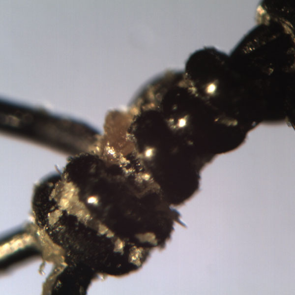
Mr. DePorter contacted McCrone Associates because he was curious if he had purchased more than just a set of used stitches. He asked to have the stitches examined under a microscope to determine if there was anything remaining that was not visible to the naked eye. Additionally, he requested some photomicrographs to include with the stitches, which are now on display at Harry Caray’s Tavern on Navy Pier in Chicago.
Solution
After the stitches arrived at McCrone Associates, they were given to our ISO 5 cleanroom staff for examination and imaging using a stereomicroscope. They were also subsequently prepared for analysis by scanning electron microscopy. Our expertise is analyzing micrometer- and nanometer-sized particles, so looking at an object that is millimeters in size presented an ironic challenge. A stereomicroscope, at a relatively low magnification, provided more than ample magnifying power to examine the stitches. Imaging the stitches, again due to their size and shape, presented a challenge because of the in-focus depth-of-field limitation of a stereomicroscope.
To accomplish the objective of providing crisp, in-focus images of an entire stitch, we utilized a scanning electron microscope (SEM). The electron optics of SEMs provide a much greater range of magnification and a much greater depth of field compared to light microscopes. However, these advantages come with the caveat that the images are grayscale.
Results
Upon examination of the stitches, it was immediately obvious that DePorter had purchased more than just a set of surgical stitches. There was some of Andrew Shaw and other debris left behind, especially in and around the knots used to close the stitches. A micrometer portion of skin was isolated and examined using a polarized light microscope. Using the polarized light microscope, which provides higher magnification and information about the composition of a material based on the interaction of light with the sample, we were able to examine and image individual skin cells.
Individually, light microscopy and scanning electron microscopy are powerful tools for the analysis and imaging of a wide variety of materials. When multiple microscopy methods are used, as they were in this case, more complete characterization of the sample under investigation can be provided.