Fractured Patterns: Microscopical Investigation of Real Physical Evidence
Conceived at McCrone Associates more than two decades ago, The Locard Exchange in its present electronic format is intended to provide a forum for the exchange of words and images devoted to the enhancement of forensic microscopy and enlightenment of the scientific, legal, and public communities who have free access to this column. Although offered by McCrone Associates, noted primarily for their polarized light microscopy of trace evidence, The Locard Exchange is open to articles about all types of physical evidence where modern microscopy of all kinds is vital. Each article will emphasize practical tools and techniques useful to accomplish the many varied tasks asked of the forensic microscopist. Illustrations and multiple images are encouraged in support of the articles. The contributions will be peer reviewed and edited in short order so acceptable articles can be posted on ModernMicroscopy.com as soon as possible.
Edmond Locard (1877-1966)
The column’s namesake, Edmond Locard, was born in France in 1877, and worked as an assistant to the forensic pioneer Alexandre Lacassagne prior to directing the forensic laboratory in Lyon, France. Dr. Locard’s techniques proved useful while with the French Secret Service during World War I, when he was able “by examining the stains on soldiers’ and prisoners’ uniforms, to determine where they had passed.” (1) He was a natural teacher, readily shared his knowledge and in his seven-volume classic Traité de criminalistique, he wrote, “To write the history of identification is to write the history of criminology”. (2) Since the time of Locard, forensic microscopists all over the world have understood that “The microscopic debris that covers our clothing and bodies are the mute witnesses, sure and faithful, of all our movements and all our encounters.” (3)
Duayne Dillon recently lamented that authors over the years have offered varying statements that they identified as “Locard’s Exchange Principle” and much has been made of the importance of this “principle.” He noted that Locard’s publications make no mention of an “Exchange Principle,” although Locard did make the observation, “Il est impossible au malfaiteur d’agir avec l’intensit que suppose l’action criminelle sans laisser des traces de son passage,” translated as “It is impossible for a criminal to act, especially considering the intensity of a crime, without leaving traces of this presence,” in the first sentence in Chapter 3 (Traces) in Manuel de Technique Policière, Paris: Payot, 1923. The first reference found of Locard’s exchange principle appears in Reginald Morrish’s, The Police and Crime-Detection Today, London: Oxford University Press, 1940, 72. (4) The Locard Exchange Principle was reiterated by L.C. Nickolls, in 1956, referencing Locard (1928), thereby confirming it as a lasting principle applicable to forensic microscopists’ work worldwide. (5) I commonly express the principle this way: whenever two objects come in contact with each other, they transfer material from one to the other. The Locard exchange produces the trace evidence of interest from fingerprints to mud.
Forensic Microscopy
Forensic microscopy is always of interest; everyone likes a good mystery; and recent high profile cases, news accounts of notable cases, and television fiction have repeatedly brought forensic microscopy into the limelight. Therefore, The Locard Exchange should be of interest to more than the scientists who use microscopy. Microscopy in various forms is used to identify and compare all types of physical evidence from all types of interesting cases. Although microscopic bits of materials in the debris and dust of our lives (hairs, fibers, paint, glass and soil) are most often thought of when we think of forensic microscopy, the other more specialized disciplines that evaluate impressions (fingerprints, bullets, footprints), fractured fragments (broken tools and torn paper), genetic markers (DNA), and handwriting also use microscopy routinely. Therefore, The Locard Exchange will be inclusive of all types of microscopy applied to all types of forensic problems. To paraphrase Locard, to write the history of forensic microscopy is to write the history of criminalistics.
Real Evidence
Microscopy leads to images that lead to real evidence. Real evidence consists of things brought into the courtroom versus the assertions of witnesses about things, and conveys a relevant firsthand sense impression to the trier of fact versus those which serve merely to report the secondhand sense impressions of others. McCormick’s Handbook of the Law of Evidence (6) provides some insight.
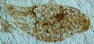
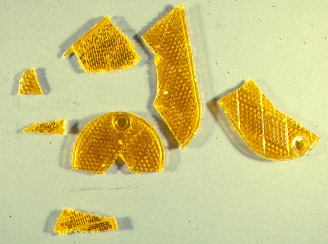
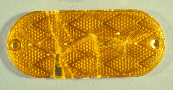
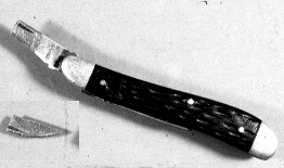
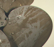
Real evidence consists of objects like guns and bullets, shoes and shoeprints [Figure 1], broken taillights [Figures 2 and 3] or broken knives [Figure 4] as distinguished from the assertions of witnesses about things. Since “seeing is believing” and real evidence with a little magnification appeals directly to the senses of the trier of fact, it is felt that this kind of evidence possesses an immediacy and reality which make it particularly persuasive. Unfortunately, real evidence remains the exception rather than the rule in many notable trials today. Most cases, particularly those cases involving miscarriages of justice, still involve the use of dubious eyewitnesses and purposeful inventions, mistaken victims, false and coerced confessions, lying jail-house snitches, inadequate defense counsel, and opinions from all types of pseudo-scientists made without scientific foundation, without scientific experiment and without real physical evidence. (7)
Real evidence possesses unusual force, and largely for this reason, real evidence is frequently objected to as “prejudicial,” because it may convey an impression of objective reality regarding an expert’s opinion. Thus the courts are frequently sensitive to the objection that the evidence is “misleading”, and zealous to insure that there shall be no advantage given the truth “in my courtroom.” Instead, they often return to the use of eyewitnesses, opinion testimony and confessions without reservation.
Apart from its bearing on the issues of the case, real evidence presents logistical difficulties for the courts. Since the courts are basically structured, architecturally and otherwise, to receive the testimony of witnesses, the presentation of real evidence may require that the court physically move to receive unwieldy objects like microscopes into the courtroom, actions which may occasionally delay and confuse the proceedings. Also, real evidence is insusceptible to a paper record for purpose of appellate review, leaving the appellate court without the benefit of the real evidence, although digital technologies should alleviate the problem.
Most forensic scientists do not seem to like the idea that the trier of fact might be allowed to make their own interpretation of the physical evidence and have, therefore, not always liked the idea of presenting the real evidence, or a photomicrograph of the real evidence, as objective proof of an association. Most have preferred to only offer their conclusions in curt reports and ultimate opinions in court. Likewise, the courts have some problems with leaving the tangible items with the jury for their consideration because of their inability to control the jury’s use of them for purposes of experimentation (doing their own analysis) outside the masterly control of the attorneys. (6)
As Paul Kirk explained at about the same time as Nickolls was explaining the Locard Exchange Principle: “Wherever he steps, whatever he touches, whatever he leaves, even unconsciously, will serve as a silent witness against him. Not only his fingerprints or his footprints, but his hair, the fibers from his clothes, the glass he breaks, the tool mark he leaves, the paint he scratches, the blood or semen he deposits or collects. All of these and more, bear mute witness against him. This is evidence that does not forget. It is not confused by the excitement of the moment. It is not absent because human witnesses are. It is factual evidence. Physical evidence cannot be wrong, it cannot perjure itself, it cannot be wholly absent. Only its interpretation can err. Only human failure to find it, study and understand it, can diminish its value.” (8) So the physical objects preserved and the microscopic images or analytical maps of real physical evidence recorded are the heart and soul of forensic science, because they, themselves, do not lie.
Fractured Patterns
Fractured patterns occur in many types of physical evidence. Most, with proper documentation, can be considered real evidence. They include the friction ridge patterns in fingerprints [Figure 5], surface striations due to tool marks [Figure 6], impressions of worn and chipped shoe soles [Figures 7 and 8], broken objects, blood spatters, and fabric impressions [Figure 9]. The probative value of these types of evidence is unlimited, fulfilling all the roles of the criminalist: discover clue materials, reconstruct events surrounding a crime, supply leads to criminal investigations, check witness statements, check confessions, demonstrate associations, strengthen other evidence, eliminate suspects, and substantiate probable guilt.
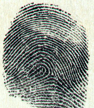

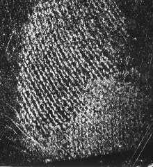
Fingerprint Microscopy
Andre Moenssens described how Locard also delved into microscopical examination of fingerprints for the purpose of using poroscopy to assist with the identifications and for the detection of fingerprint forgeries. Locard went beyond the variations of the individual fingerprint patterns which Sir Francis Galton noted as he defined the friction ridge events. Locard concluded and proposed, in the first decade of the 20th century, that identity could be established by a comparison of the pore structure in an unknown latent print with that of the inked impression. He discovered that there is an almost limitless variety of shapes and locations of pores. He contended that poroscopy was an ideal method for identifying individuals where only an infinitesimal portion of friction ridge skin was available in the latent print. Perspiration which forms part of the latent fingerprint flows to the surface of friction ridges through pore openings which are lined up along the ridges. In 1912, Locard demonstrated the value of poroscopy in the now famous case of Boudet and Simonin, where he showed the concurrence of 901 separate sweat pores in Boudet’s latent fingerprint on a piece of furniture at the crime scene and more than 2,000 in a latent impression left by Simonin’s palm.
The forgery of fingerprints was also investigated by Locard. In 1907 he discovered a procedure for making counterfeit fingerprints by the use of a mold (like a rubber stamp) made from gutta-percha. There are two relatively easy ways to forge fingerprints. One is to make a stamped print which is a replication of a fingerprint by a rubber stamp, cast, molding, plate, or die, i.e., a reproduction. The other is to collect an original latent fingerprint from an innocent surface and claim it was collected from another incriminating surface, i.e., a transferred powdered lift. The telltale ragged edging and ghost edges of stamps are diagnostic and allow the determination of a fake. The transferred powdered lift can often be associated by ultramicroanalysis of the microscopic debris in the lift with its original location based on the Locard Exchange Principle. (9)
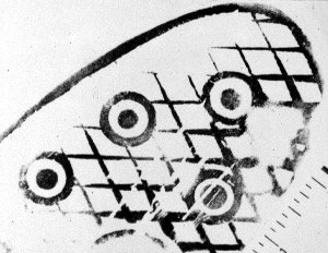
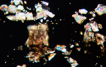
A little more recent experience confirms Locard’s methods. “True dust is the microscopic debris which is a disintegration product of our environment or structures. In this case, a fingerprint was developed on a windowsill and subsequently identified as the defendant’s. The following question was posed: By an analysis of the dust collected on the tape used to lift the latent fingerprint, could it be determined from where that print was collected? It was postulated that the lift may indeed have been collected from another place. The windowsill was marble and the other possible surface was painted. Portions of the transparent tape lift were separated from its paper backing and placed sticky side up on a microscope slide, covered with a cover slip, and cleared with xylene. No paint particles were found anywhere on the tape, but in a potion of the tape where black fingerprint powder had apparently accumulated in a crack in the surface, CaCO3 crystals were found in abundance, similar in all respects to marble dust.” [Figure 10] (10)
Firearms Microscopy
The firearm from which a bullet or cartridge case has been fired is identified by the microscopical comparison of the unique striae (toolmarks) left on the bullet or cartridge case from the worn, machined metal of the barrel, breach block, extractor, or firing pin in the gun. The history of forensic firearms and toolmark identification really coincides with the history of the comparison microscope. It began with the collaboration of Charles E. Waite, a para-legal, Max Poser of Bausch and Lomb Optical Company, John H. Fisher, a physicist, Phillip O. Gravelle, a chemist, and Calvin Goddard, a medical doctor from Johns Hopkins Hospital. It was Gravelle who mistrusted his memory. “As long as he could inspect only one bullet at a time with his microscope, and had to keep the picture of it in his memory until he placed the comparison bullet under the microscope, scientific precision could not be attained. He invented the comparison microscope and Goddard made it work.” (11)
Sir Sydney Smith, in collaboration with A. Lucas in Cairo, brought the comparison microscope to Scotland almost immediately and introduced forensic firearms identification to the European scientists. He also emphasized the importance of an initial examination of the bullet with a stereomicroscope and the value of fabric impressions and microtraces on the questioned bullet as predicted by the Locard Exchange Principle. (12) For example, paint on a bullet may demonstrate from which surface the bullet ricocheted [Figures 11 and 12].


The analysis of gunshot residues (GSR) using scanning electron microscopy (SEM) has proven to be a valuable addition to the trace evidence arsenal. The basic principle upon which the technique depends is that small particles characteristically left as a residue after a weapon is fired can be collected using adhesive-coated SEM stubs. The subsequent analysis is performed in the SEM using energy dispersive x-ray spectrometry (EDS) directly on the adhesive surface and particles unique to GSR are identified by their morphology and chemical composition. [Figure 13] Particles containing lead, antimony and barium are considered unique to gunshot residues and are real microscopic evidence associated with the firing of a gun.
Fragment Fracture Matching
Fragments are broken or torn objects, split apart by force (fractured) into separate pieces, for example, a broken tool or a torn piece of lunch bag. If one piece is found at the crime scene and another matching piece in the perpetrator’s pocket, an association is established. The types of physical matches are classified as: (1) fractured in planes with two dimensional irregular edges or cuts through unique arrays, or (2) fractured solids with either two contiguous surfaces or closely spaced non-contiguous surfaces. The match criteria are: (1) pieces must have been fractured, (2) pieces can be realigned, (3) pieces must interfit along an irregular edge-to-edge border, verified by surface markings, or verified by a three-dimensional fit, and (4) pieces must possess accidental characteristics, or unique design. Positive identifications are accomplished with magnified observation of the fractured surfaces and refitting, and made real evidence by photomicrography.
Conclusion
These illustrations of forensic microscopy of real evidence are intended as an invitation to join The Locard Exchange. Forensic microscopy not only includes trace evidence analysis (hairs, fibers, paint, glass and soil) where polarized light microscopy (PLM) is king; but other methods are of equal value: the hand lens (or camera macro lens) for fingerprints and footwear impressions; stereomicroscope for marijuana identification; infrared microscope for drug identification; phase contrast microscope for glass comparisons and the identification of spermatozoa; comparison macroscopes (as Leitz describes them) for bullets, cartridge casings, and toolmark examinations; the transmitted light comparison microscope for hairs and fibers; the scanning electron microscope for gunshot residues, and the transmission electron microscope for paint pigments. The forum is open and inclusive including articles and letters to the editor, if you would like to comment on what has been written and presented in the column.
References
1. H. Ashton-Wolfe, The Forgotten Clue: Tales and Methods of the Sûrete. London: Hurst & Blackett, 1930. Page 26.
2. La petite histoirede la médecine légale à Lyon, http://web.lyon.iufm.fr/formateurs/lyon/product/policesc/legale/medleg4.html.
3. Edmond Locard, “The Analysis of Dust Traces,” The American Journal of Police Science, Volume 1, 1930, Page 276.
4. Duayne J. Dillon, D. Crim., http://groups.yahoo.com/group/forensic-science/message/2857, Tuesday, July 9, 2002, 5:16 p.m.
5. L.C. Nickolls, The Scientific Investigation of Crime. London: Butterworth & Co., 1956. Page 39.
6. Edward W. Cleary, editor, McCormick’s Handbook of the Law of Evidence. St. Paul: West Publishing Co., 1972. Pages 524-542.
7. Jim Dwyer, Peter Neufeld, and Barry Scheck, Actual Innocence. Signet Books. New York, 2001.
8. Paul L. Kirk, Crime Investigation. New York: Interscience Publishers, 1953. Page 4.
9. Andre A. Moenssens, Fingerprint Techniques. Radnor, PA: Chilton Book Company, 1971.
10. Richard E. Bisbing, “Clues in the Dust,” American Laboratory, November, 1989.
11. Jurgen Thorwald, The Century of the Detective. New York: Harcourt, Brace & World, 1964.
12. Sir Sidney Smith, Mostly Murder. London: George G. Harrap & Co, 1959.
Comments
add comment