Microscope Activities, 9: Calibrating the Field of View
In the past, Hooke College of Applied Sciences offered a microscopy workshop for middle school and high school science teachers. We thought that these basic microscope techniques would be of interest not only for science teachers, but also for homeschoolers and amateur microscopists. The activities were originally designed for a Boreal/Motic monocular microscope, but the Discussion and Task sections are transferable to most microscopes. You may complete these 36 activities in consecutive order as presented in the original classroom workshop, or skip around to those you find interesting or helpful. We hope you will find these online microscope activities valuable.
EXPERIMENT 9: Calibrating the Field of View
Goal
To determine the diameter of the field of view for each objective supplied.
Level
Basic
Materials Needed
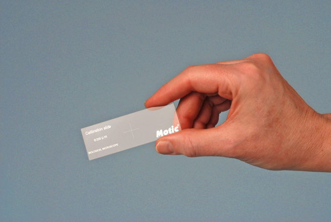
- Motic Calibration Slide (supplied).
- Optional: stage micrometer; transparent metric rule
Procedure
Supplied with the Boreal/Motic microscope is a calibration slide which consists of a metalized dot 200 μm in diameter, at the center of interrupted crosslines (Figure 9-1).
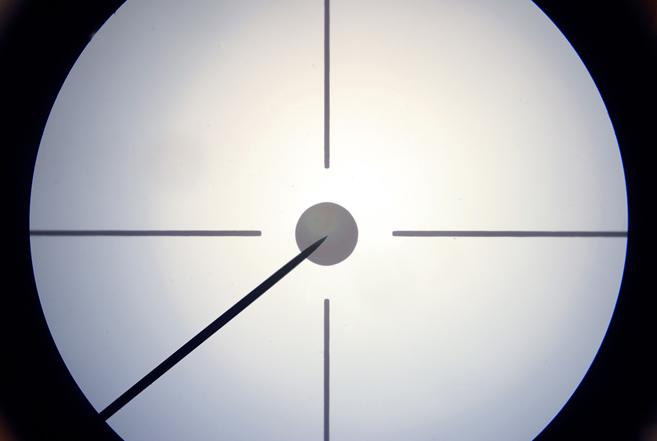
Viewing the calibration slide with each objective in turn, determine the diameter of the field of view; the use of the mechanical stage vernier may be helpful here. Figure 9-2 is the view of the calibration slide using the 10X objective. The diameter of the central calibration disc is 200 μm; the eyepiece pointer is also visible.
As a matter of interest, you may also want to focus on a transparent metric rule, using each objective.
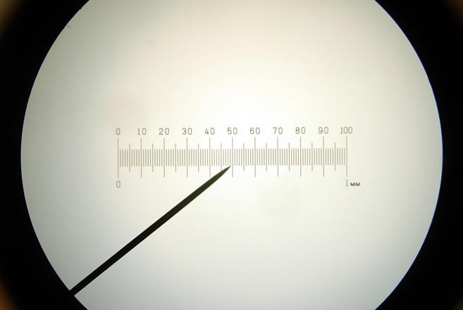
A more accurate determination of the field of view diameter may be gained by using a stage micrometer, if one is available. Figure 9-3 is the view of a 1 mm stage micrometer divided into 100 parts as seen using the 10X objective; a 2 mm or 5 mm stage micrometer would save a little time, because the longer scales would cover the entire field of view, even with the 4X objective.
It is curious that a 200 μm diameter disc and crosslines calibration slide would be supplied, when it would not have been more expensive or troublesome to provide a micrometer scale.
Discussion
Knowing the diameter of the field of view for each objective is vital for estimating the size of microscopic specimens, the growth rate of crystals, and the velocity of moving specimens. Of course, a calibrated eyepiece micrometer would be even better, and this is why we advocate loosening/removing the eyepiece lock screw to allow other eyepieces to be used.
Teacher’s Note
Using a transparent metric rule, the diameter of the field of view can be crudely estimated to be 4X ~ 4.5 mm, 10X: <2 mm. 40X: < 1 mm.
Using a stage micrometer, the diameter of the field of view with our microscope was measured to be:
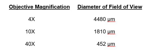
These values are important and used frequently, so that it is a good idea to write these values on a microscope slide label, and attach the label somewhere on the base of the microscope stand so that it can be consulted quickly when needed.
Hint: If you have a stage micrometer, it would also be a good idea to determine the width of the eyepiece pointer, using each objective, as an aid in estimating the size of microscopic specimens and structures. With our microscope, we determined the width of the eyepiece pointer to be:
4X = 66 μm
10X = 30 μm
40X = 8 μm
Again, these values should be recorded on a slide label, and affixed somewhere on the base of the microscope stand.
We also determined the actual diameter of the supplied 200 μm calibration disc to be 196 μm.
Stage micrometers are expensive (~$150) if purchased new. Used stage micrometers are available through online auctions, with starting bids of $1.50 to $49. New, imported stage micrometers are available for $40-$50. Stage micrometers are only needed once to calibrate eyepiece micrometers and field-of-view diameters, so that only one shared stage micrometer is needed per school or district.
Task
Determine the diameter of the field of view for each objective, as described in the Procedure section; record the values on a slide label, and attach the label to the base of your microscope. If you have an eyepiece micrometer this would be the time to calibrate it against a stage micrometer.
Comments
add comment