Julian D. Corrington and the Bausch & Lomb Microscopes for Amateurs
The name Julian D. Corrington, and Bausch & Lomb’s Model R, Gem, and New Gem microscopes for young people and other beginners are forever linked in the history of amateur microscopy in America. Indeed, Corrington has been called “the father of amateur microscopy in the US” (Santacana 2000, 2012). New users of the microscope seeking information through books from the library are likely to find at least one of Corrington’s three main books on the microscope for beginners: Adventures with the Microscope, Working with the Microscope, and Exploring with Your Microscope. All of these books feature Bausch & Lomb microscopes made specifically for amateurs and other beginners. How this all came about is the subject of this story.
Fortunately, we have Corrington’s biography as told in his own words. We’ll let him tell his story, and then we will fill in the gaps. The New York Microscopical Society (NYMS) celebrated its centennial in 1977. In the year following the New York Microscopical Society Centennial, Hal Bowser, who was editor of the NYMS’s excellent newsletter, invited Corrington, who was then living in retirement in Florida, to update the membership through a “Greetings from an Old Friend.” In his response, Corrington, after extending congratulations to the NYMS as an organization on their centennial, had this to say:
“In a recent letter from Hall Bowser he suggested that I might tell the older members what has become of one who, in his words, ‘practically invented amateur microscopy,’ indeed a flattering compliment. At the time of my first articles on this subject I was, at age 40, head of the microscope slide department and editor of their Bulletin and catalogs for Ward’s Natural Science Establishment at Rochester, N.Y. I had written my Ph.D. thesis in zoology, at Cornell University, and had produced a few scientific papers, but I was by no means what could be called a ‘writer.’ Then the Bausch & Lomb Optical Company asked me to do some booklets to accompany sales of their small New Gem and later Model R microscopes for young people and other amateurs and beginners. These booklets proved so successful that finally they authorized me to do a full-sized book (455 pages) Adventures with the Microscope, 1934. As I was under full-time employment at Ward’s, I had to write and rewrite and rewrite this large volume nights, weekends, and holidays. But at length it was done, and I had learned to write by writing—and that’s the best advice any aspiring author could receive.
“Since that time my bibliography shows a total of 450 publications, including eight of book length, the best known being McGraw-Hill volumes, Working with the Microscope, 1941 and Exploring with Your Microscope, 1957, the latter also having a Serbo-Croat translation published in Beograd in 1972. My longest series of monthly articles ran in Modern Mechanix (later Mechanix Illustrated), Leisure, Practical Microscopy, Bios, and above all Nature Magazine, to which I was a contributor for 22 years. These years also saw the formation of the American Society of Amateur Microscopy (A.S.A.M.) for which I did thousands of hours of dogged but very rewarding labor.
“Before coming to Rochester I had, like the great majority of people, associated the microscope only with the biological laboratory in high school, college, medical schools and hospitals, but my connections with Ward’s and Bausch & Lomb soon disabused me of such a narrow view. For magnifying instruments of many types have a widespread use in industries of many sorts—paper, textiles, paints, glass, rubber, leather, and so on through a long list. And so my aim in all of these series of books and magazine articles has been to start the beginner off with easily prepared slides: I advise them to mount a postage stamp, a piece of newsprint, a small square of silk or cotton cloth clipped from the hem of a dress, grains of sand, a fingerprint—and only later to take up the much more complicated procedures of fixing, sectioning, mounting and staining of, say, a section of frog liver or cat intestine. I urge newcomers to microscopy to start with fundamentals and build up gradually into the numberless fields of use for the microscope. To this end, I employ simple language, hoping that the beginner will establish a solid foundation for his or her later specialized work. It is for this system and approach that I believe I can claim any originality in my writings.
“In 1944, I came to the University of Miami, where I have taught mainly premedical courses—comparative anatomy, histology, and embryology—retiring in 1962. Now, at age 86 I am enjoying senior citizen leisure, and am happy in the fact that many former students, readers, and friends tell me that I have been of help to them in their careers.”
Biography
Ancestry records show that Julian Dana Corrington (1891 – 1979) was born in Arkansas on December 22, 1891 to John W. Corrington (1861 – 1945) and Julia Knox (1875 – unknown). He had a U.S. World War I Draft Registration Card (1917 – 1918) while a resident in Tompkins, New York, and a U.S. World War II Draft Registration Card (1942) while a resident in Maryland, but there is no record of his having served in the military. The 1920 United States Federal Census lists his spouse as Florence H. Corrington, and their residence as Tompkins, New York; the 1930 Census lists their residence as Onondaga, New York.
In his autobiographical sketch to the NYMS, Corrington mentions that at age 40 (1931) he was working for Ward’s, and that he had already done his Ph.D. in zoology (Cornell University), but he had already published a number of articles typical of zoologists at the time, and had been teaching zoology and botany since 1926. An example of his publications at the time include, “The Winter Birds of the Biloxi, Mississippi Region” (Corrington 1922), in which he describes 112 species of birds which he observed during the winter of 1920 – 1921; he was a member of the Department of Zoology at Cornell University at the time. Other publications included “Commensal Association of a Spider Crab and a Medusa” (Corrington 1927), “Morphology of the Anterior Arteries of Sharks” (Corrington 1930), and “Abnormal Herpetological Specimens from Syracuse” (Corrington 1931). A note in the journal Science 54 329 October 7, 1921 states that Corrington resigned as curator in the Department of Zoology of Cornell University to accept the appointment of associate professor of biology at the University of South Carolina, Columbia, South Carolina.
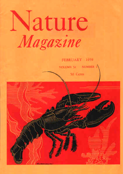
In addition to the professional articles mentioned above, which we would typically expect from a Ph.D. zoologist, are Corrington’s contributions to the more general interest magazines he casually mentions in his autobiography. One of the most popular of these is Nature Magazine (Figure 1). This magazine had a 36-year run of 52 volumes from 1923 to 1959, and Corrington wrote and edited the column “Under the Microscope” for this magazine for 22 years. It was the popularity of these columns, written for amateurs for almost a quarter of a century, that brought him to the attention of Bausch & Lomb when they were looking for an author of booklets to accompany their microscopes made for beginners. The great interest shown in amateur microscopy led Bausch & Lomb to invite Corrington to write a full-size book on microscopy for beginners that would, of course, feature Bausch & Lomb microscopes.
In 1939, Corrington founded the American Society of Amateur Microscopists (ASAM), and issued a certification to its members that was signed by its founder.
Publications on the Microscope
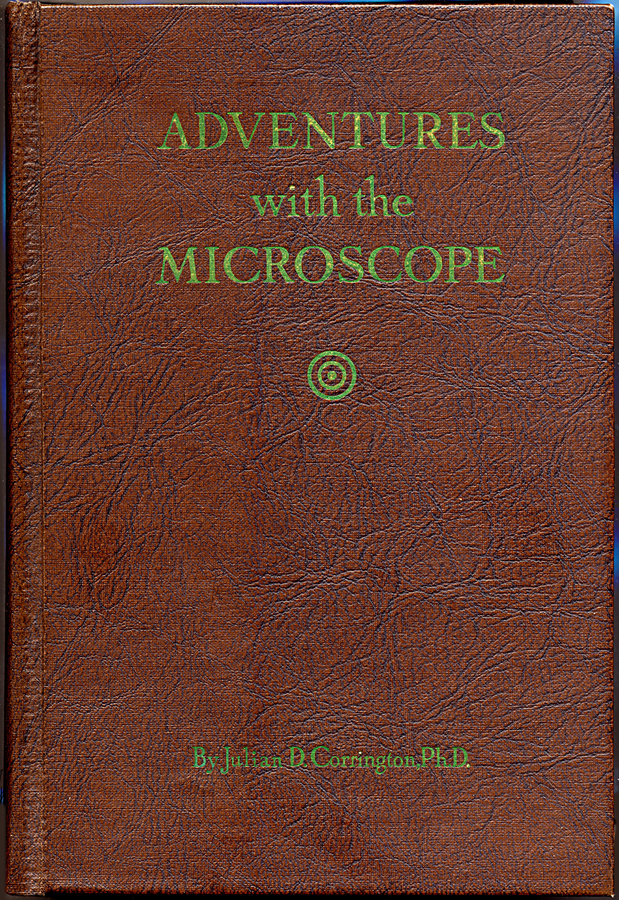
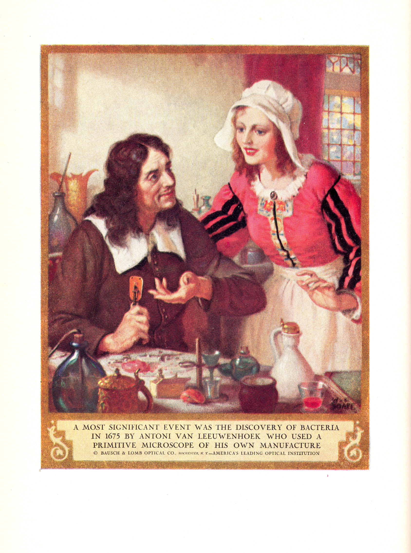
Corrington’s Adventures with the Microscope (Corrington 1934) was written while he was still working for Ward’s, as indicated on the title page (Figure 2). The 455-page book had a leather-look cover (Figure 3), and the frontispiece featured one of the color plates (Figure 4) from Bausch & Lomb’s series of paintings on the history of optics. This important publication has descriptions and illustrations of Bausch & Lomb’s New Gem microscope and the Model R microscope, as well as the optional photomicrographic apparatus being sold for the Model R.

For many readers, one of the most favorite chapters in Adventures with the Microscope is the last chapter, “Sherlock Holmes Buys a Microscope;” the chapter is in a narrative form, as related by Dr. Watson. A number of Bausch & Lomb microscopes are illustrated here, including the Model FFSB student monocular microscope, the Comparison Microscope (with and without the photomicrographic apparatus), and the AKW-5 Wide-Field Binocular Microscope. This chapter also illustrates a fingerprint card, and gives an introduction to fingerprint identifying characteristics and fingerprint matching. (As an aside, it is interesting to note that the photos for Adventures with the Microscope were made by Charles S. Foster, Eastman Kodak Company, who would also make contributions to microscopy.)
Corrington’s next book, Working with the Microscope (Figure 5), was published in 1941 (Corrington 1941). The microscope shown being used on the dust jacket is not one of the “baby” microscopes, but appears to be the Model H Professional Laboratory Microscope, which in 1938 was being sold for $83.00. This book went through at least seven printings.
By 1957, Adventures with the Microscope had already been out of print for a number of years, and so it was deemed advisable to come out with a new work which, in general plan, was to be an abridged and revised version of the 1934 work. Some of the material and illustrations were taken from the more technical Working with the Microscope (1941), but the bulk of the new book, Exploring with Your Microscope (Corrington 1957) (Figure 6), was new in both scope and purpose. The last chapter in Exploring with Your Microscope parallels the last chapter in Adventures with the Microscope in that it is titled “Adventuring with Sherlock Holmes.” The chapter opens with a quotation from Arthur Conan Doyle’s “The Adventure of Shoscombe Old Place”; specifically, where Sherlock Holmes “had been bending for a long time over a low-power microscope,” and then invites Watson to observe the glue, epithelial “scales,” dust, and threads from a tweed coat. A wonderfully appropriate photomacrograph at 11X is provided to illustrate the field of view. The fingerprint information is also here, but the comparison microscope illustration has been updated (as has the microscope on the dust jacket). There is also a photomicrograph of a bat hair (741X).
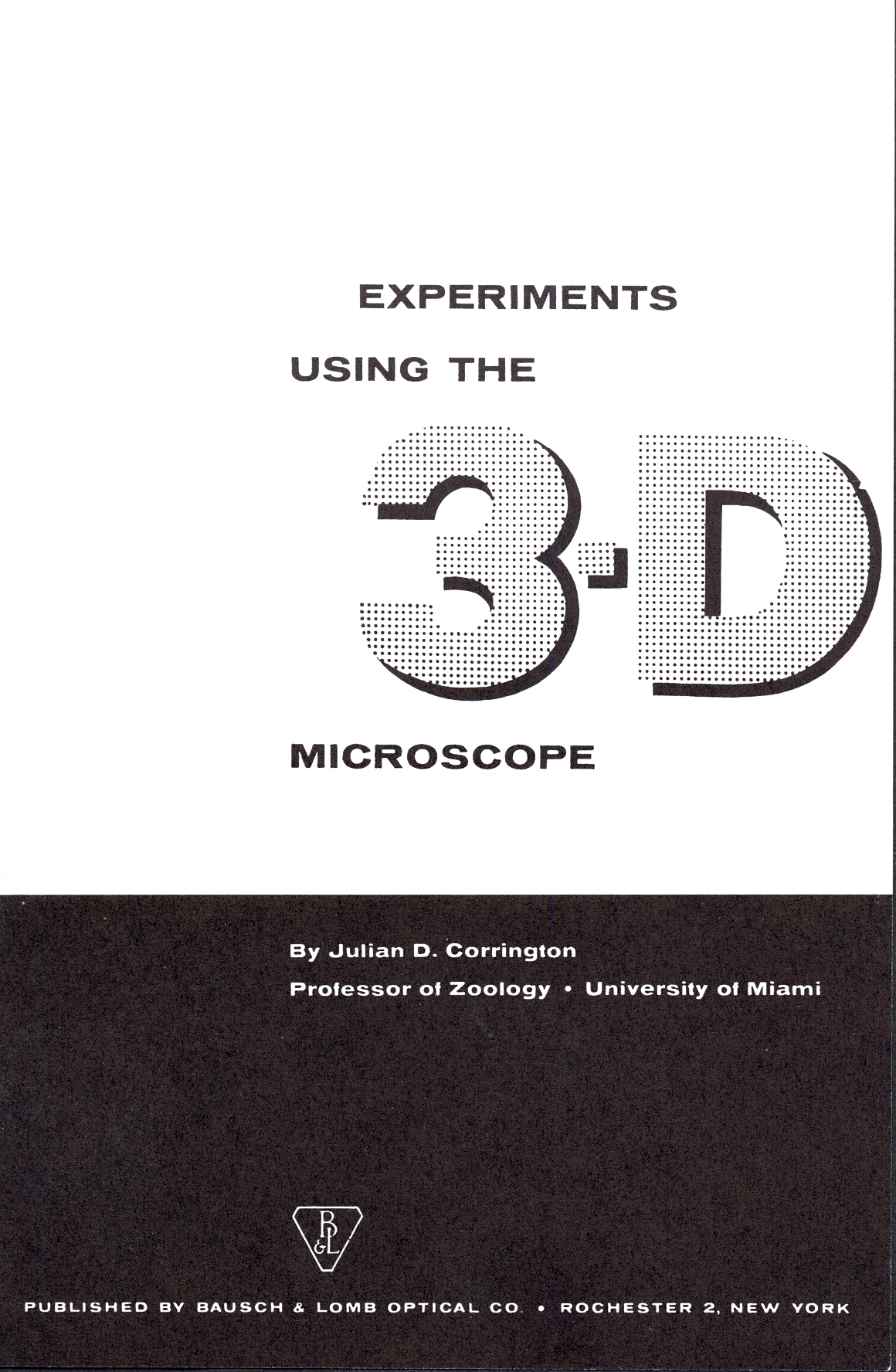
In addition to these three full-length books written for amateur and advanced amateur microscope users, Corrington was writing various booklets. One of these booklets, written after he had already moved to Florida in 1944 to teach at the University of Miami was Experiments Using the 3-D Microscope (Corrington 1959). This publication (Figure 7) was written on behalf of Bausch & Lomb.
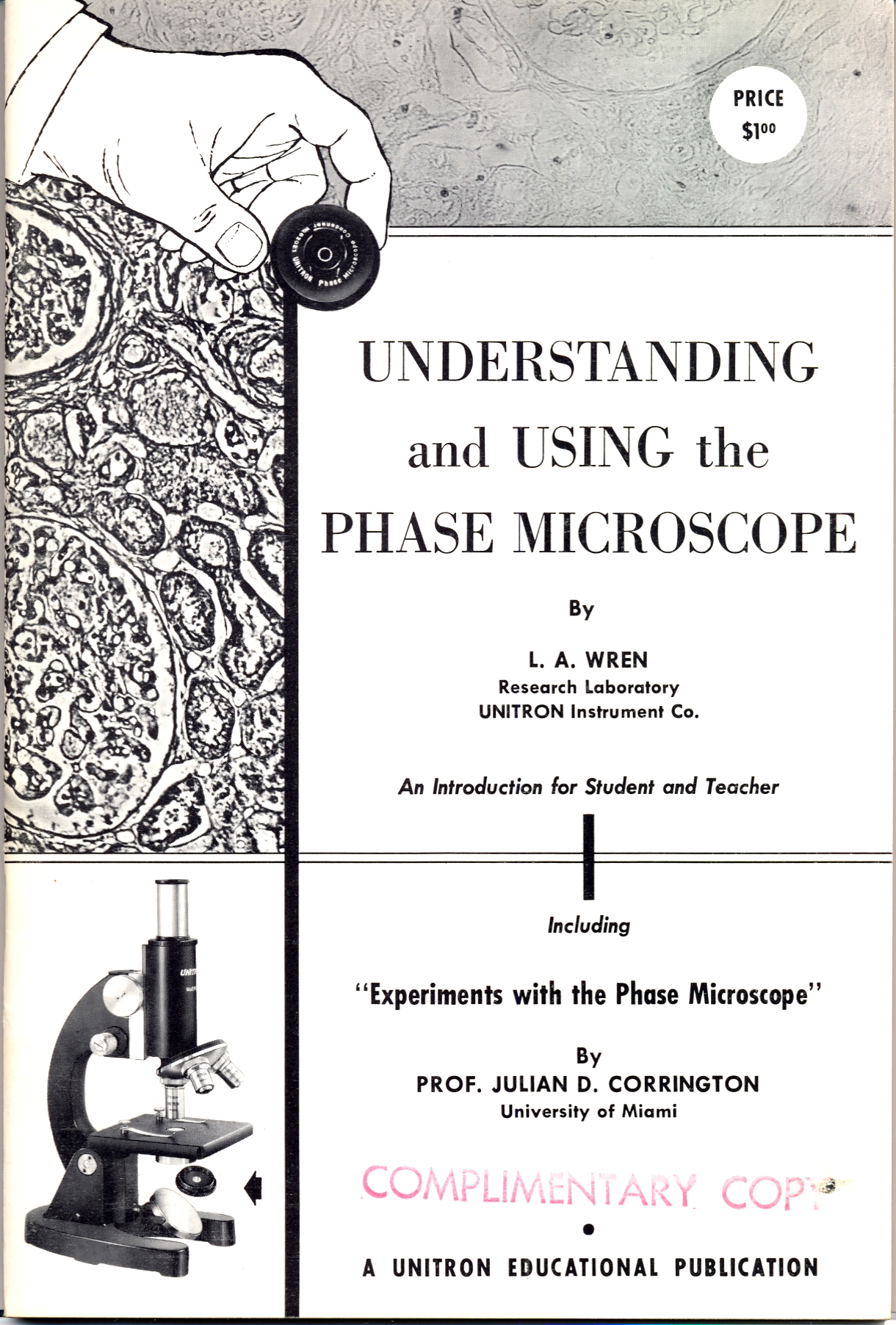
Even after retiring from the University of Miami in 1962, Corrington’s services were still being sought. When the Unitron Instrument Co. Had one of their research laboratory personnel, L.A. Wren, write a booklet on Understanding and Using the Phase Microscope (Wren 1963), Corrington was asked to be included by writing a series of Experiments with the Phase Microscope (Figure 8).


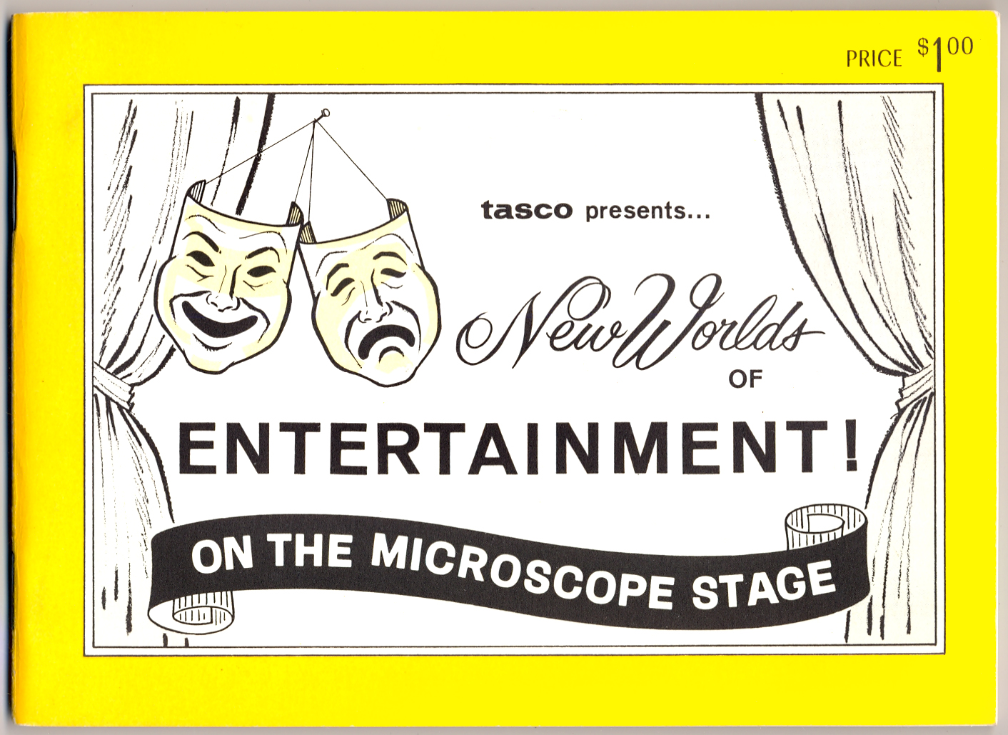
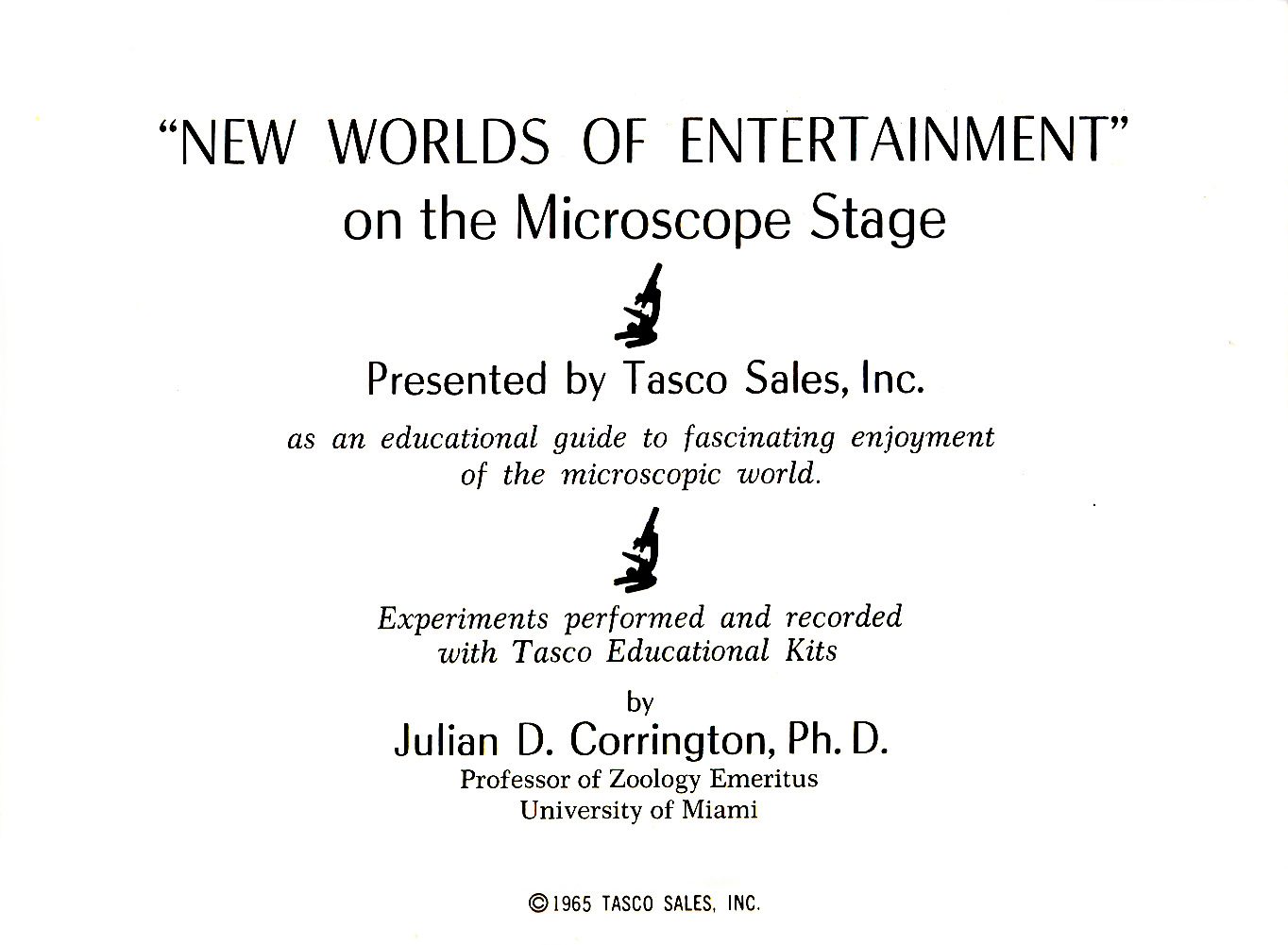
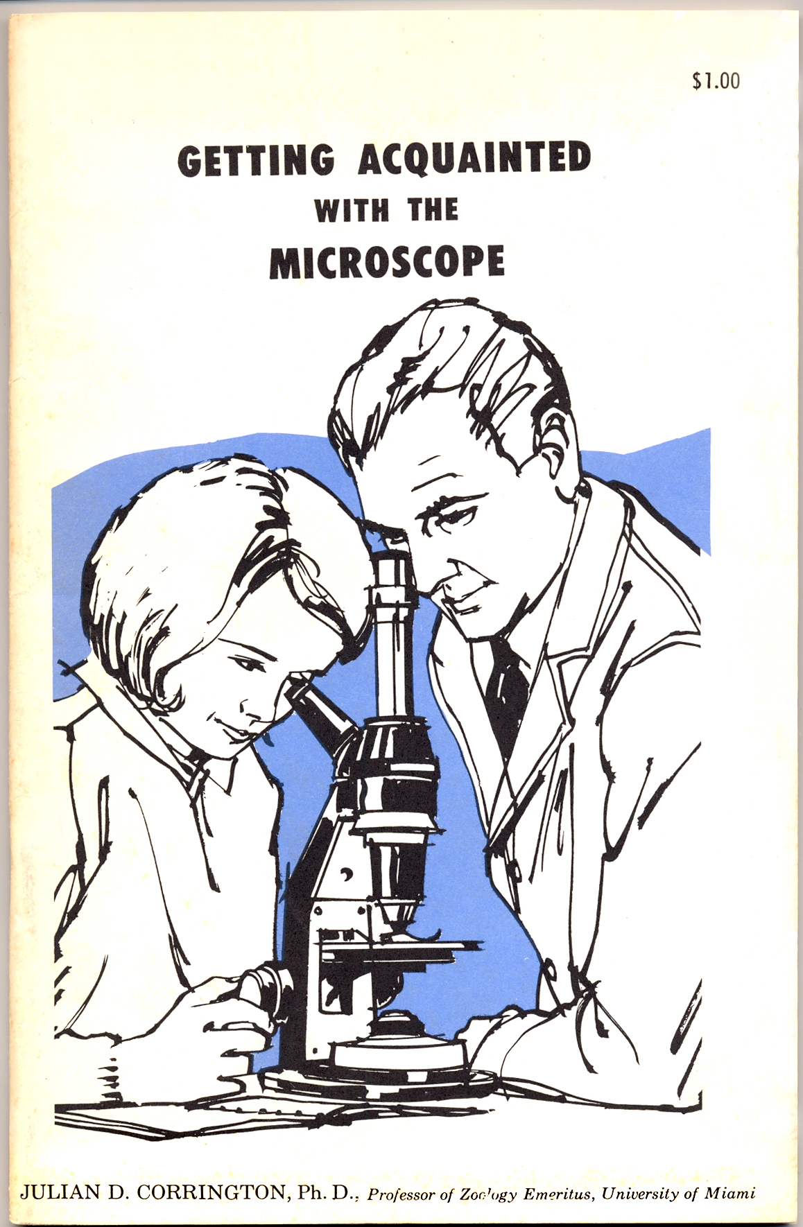
In 1969, seven years after retiring, Corrington wrote yet another booklet for Bausch & Lomb (Corrington 1965); this one was called Getting Acquainted with the Microscope (Figure 11).When, in 1965, Tasco Sales, Inc. put together a rather extensive microscope set featuring their microscope for amateurs (Santacana 2000), they asked Corrington to write the manual (Figure 9) accompanying the set. Corrington wrote New Worlds of Entertainment on the Microscope Stage (Corrington 1965) as Professor of Zoology Emeritus (Figure 10).
The Bausch & Lomb Microscopes for Amateurs
The principal microscopes for amateurs offered by Bausch & Lomb throughout the 1930s are the Model R and the New Gem, both of which are featured in Adventures with the Microscope. The Model R microscope is today scarce because during the early 1930s depression years, when the microscope sold for $21.00, with a copy of Adventures with the Microscope, not many people could afford it, and by the end of the decade, Bausch & Lomb had already converted over to the manufacture of binoculars and other optical equipment for the military; World War II had already started in Europe. Bausch & Lomb never resumed the manufacture of the Model R after the War. Some idea of the difficulty in finding a Model R today is indicated by the title of an article on the microscope written by Charles Gellis for the Journal of the Microscope Historical Society: “In Pursuit of the Elusive Model R Microscope: Observations and Measurements” (Gellis 1998).
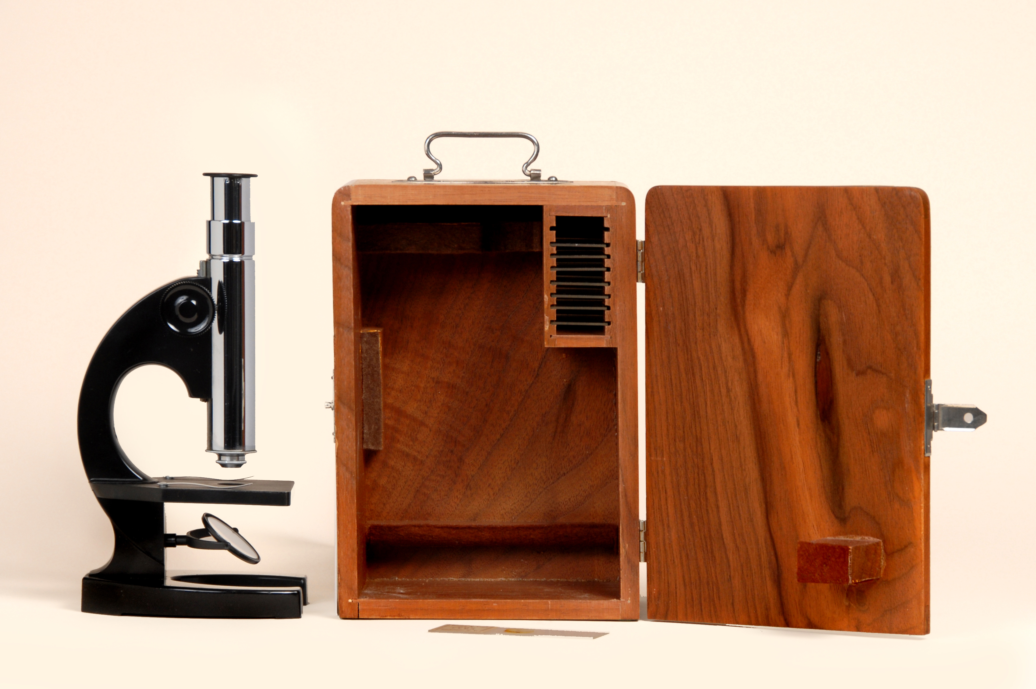
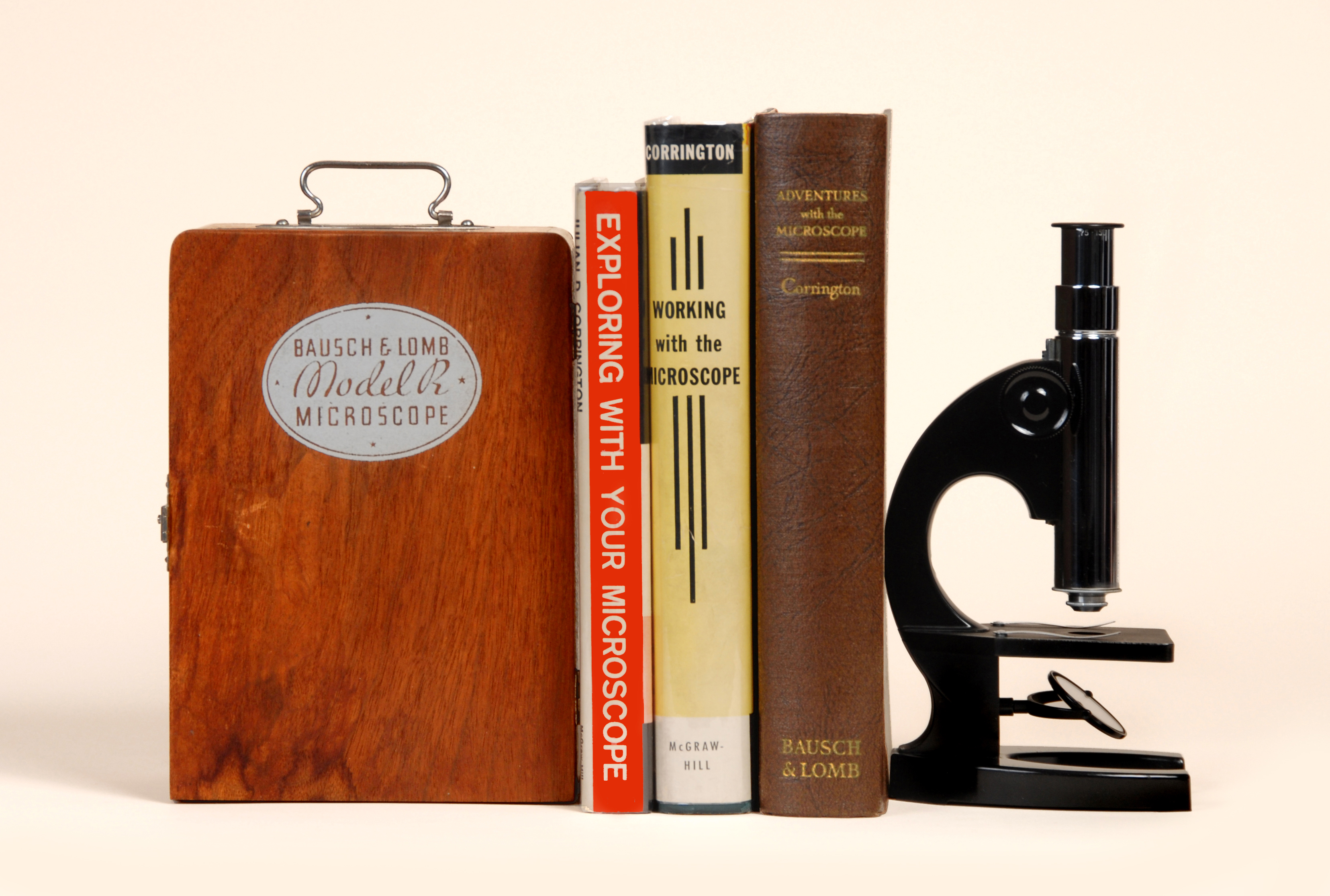
The Model R microscope came in a beautiful walnut carrying case (Figure 12), together with a few blank slides and a prepared slide of a flea—the preparation featured in Adventures with the Microscope. The front door of the walnut carrying case had a stenciled label giving the model and manufacturer’s name (Figure 13, illustrating, also, the spines of Corrington’s books).
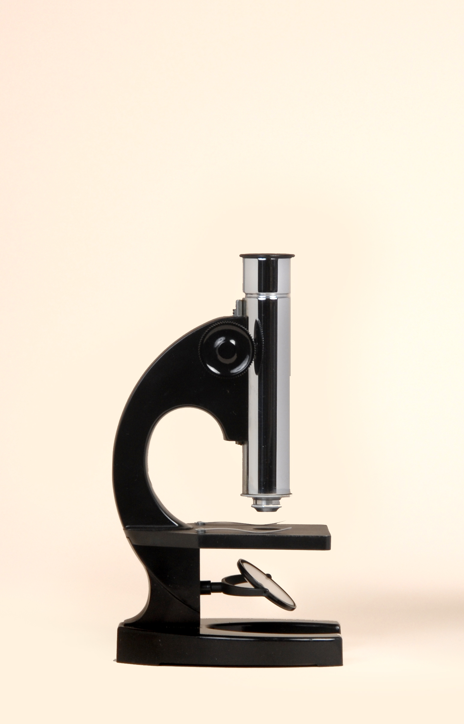
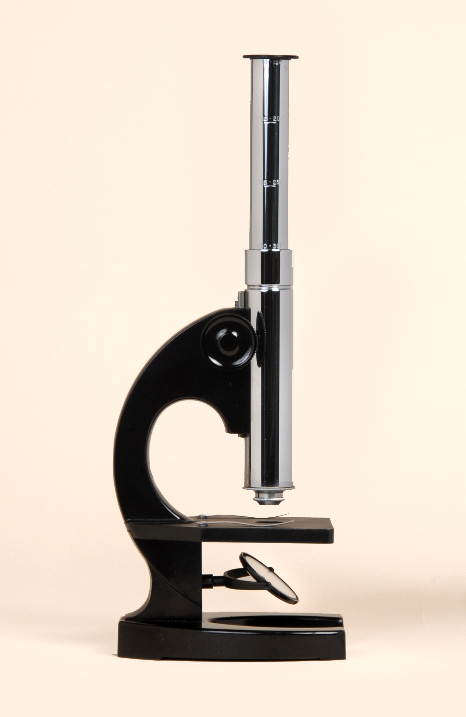
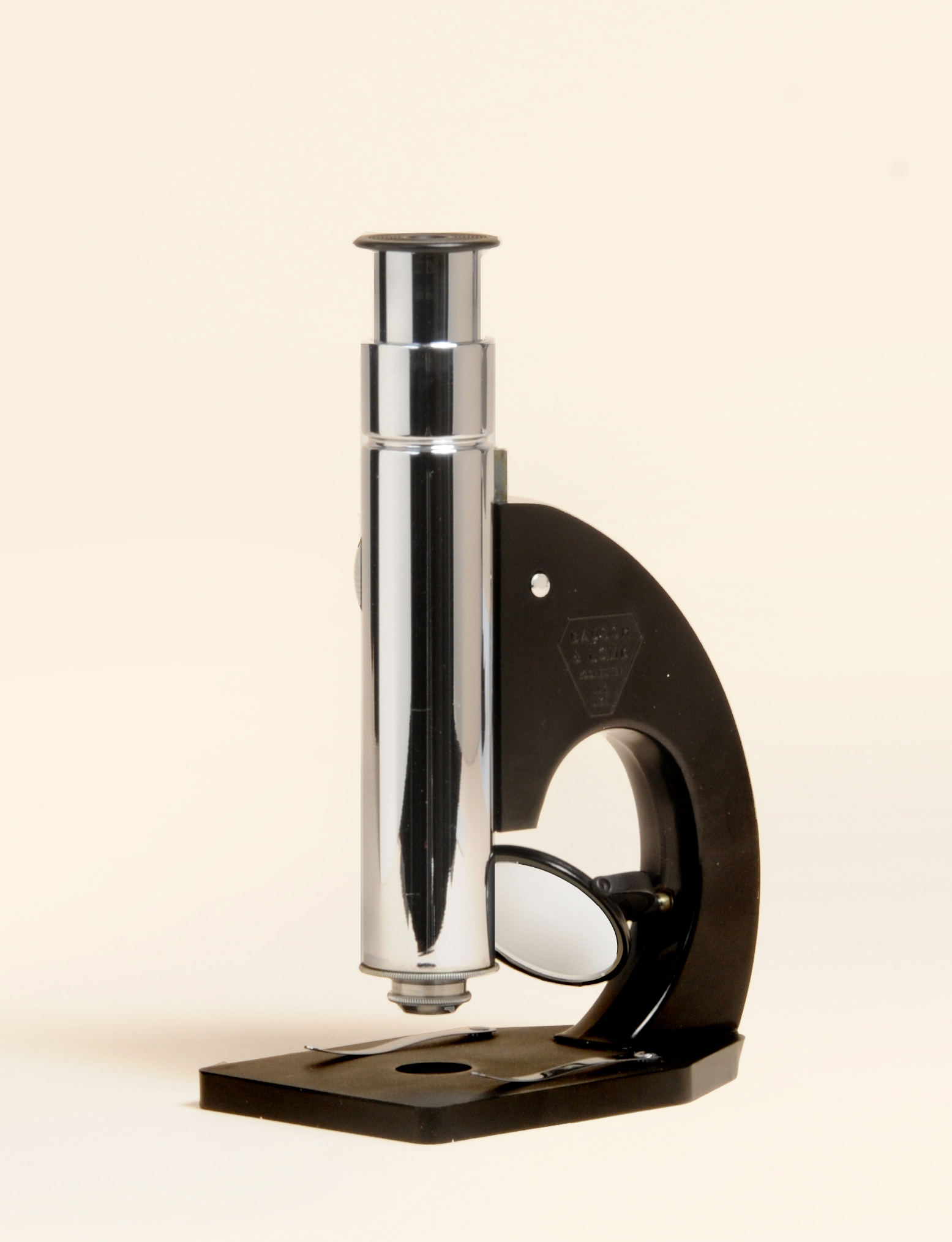
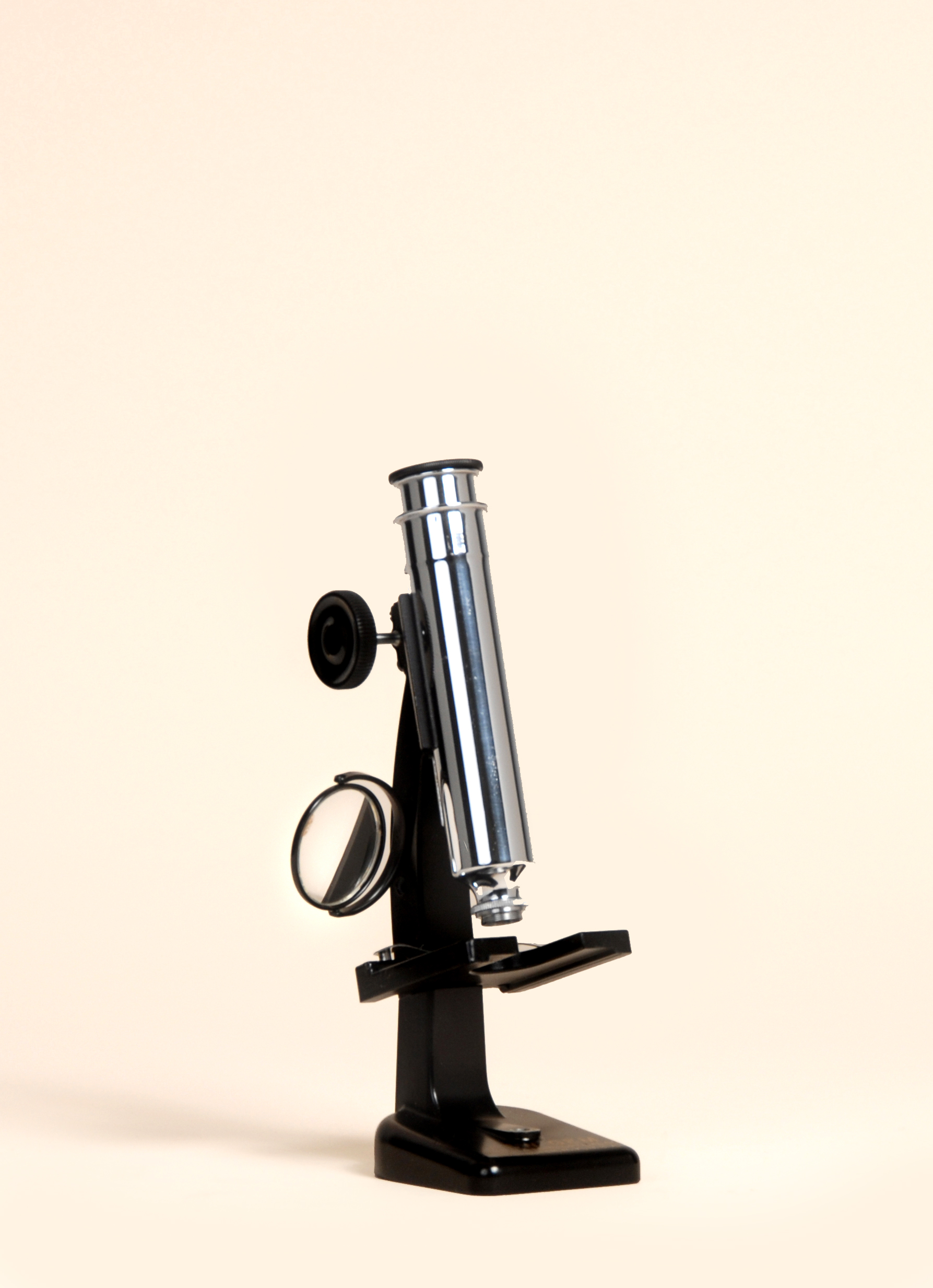
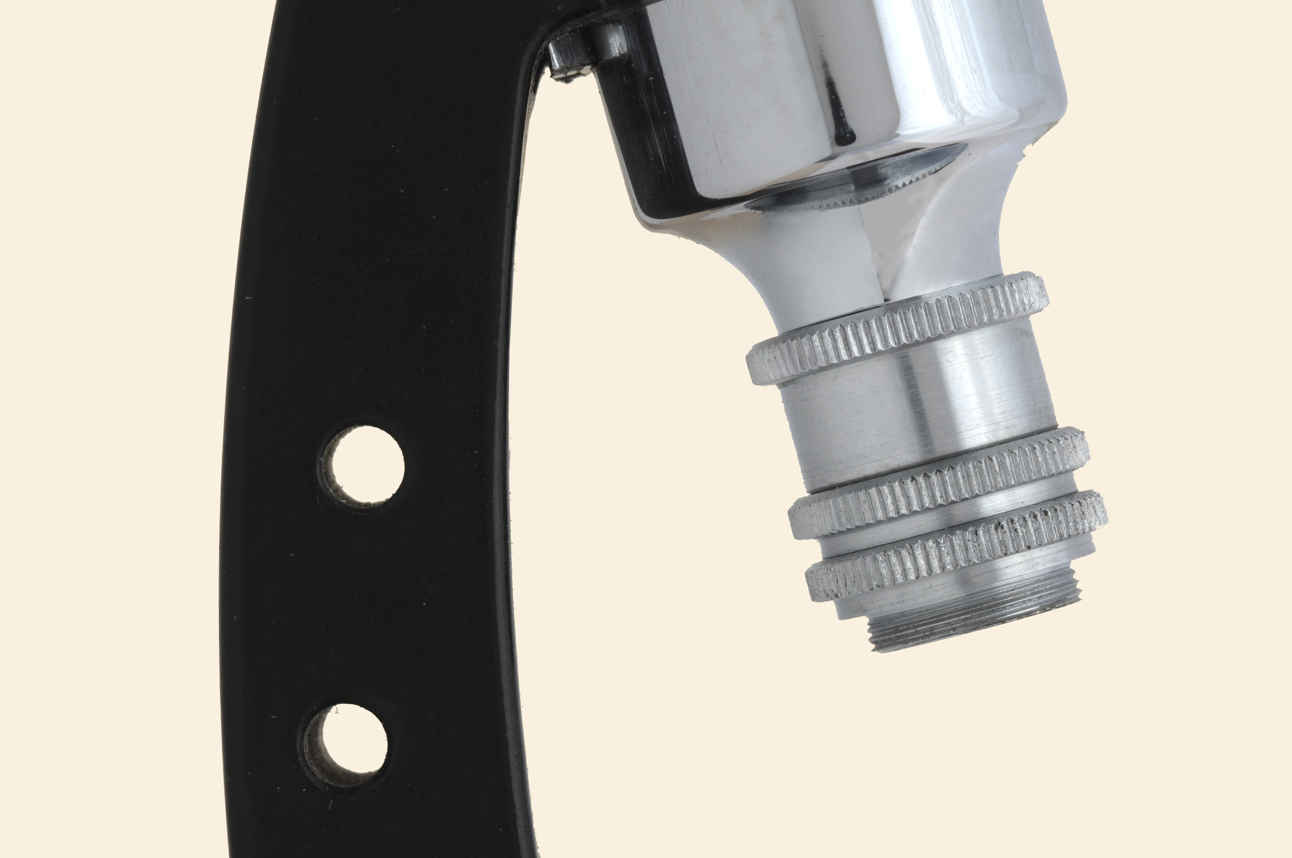
The Model R microscope itself was capable of a range of magnifications through use of a 3-1/4 inch drawtube, a divisible objective, and an optional low-power eyepiece substituted for the standard 10X. With fully-closed drawtube (Figure 14), the magnification range is 75X – 150X, depending on the objective configuration; with one-inch drawtube extension the magnification range is 125X – 250X; with fully-extended drawtube (Figure 15) the magnification range is 150X – 300X; the magnification ranges are indicated on the chromium-plated drawtube. Fully extended, the body tube is 7-3/8 inches. The image quality with drawtube fully extended is not very good because the maximum useful magnification based on the unstated numerical aperture of the compound objective is exceeded, but use of the lower-magnification eyepiece improves the image quality, making the excess less evident.
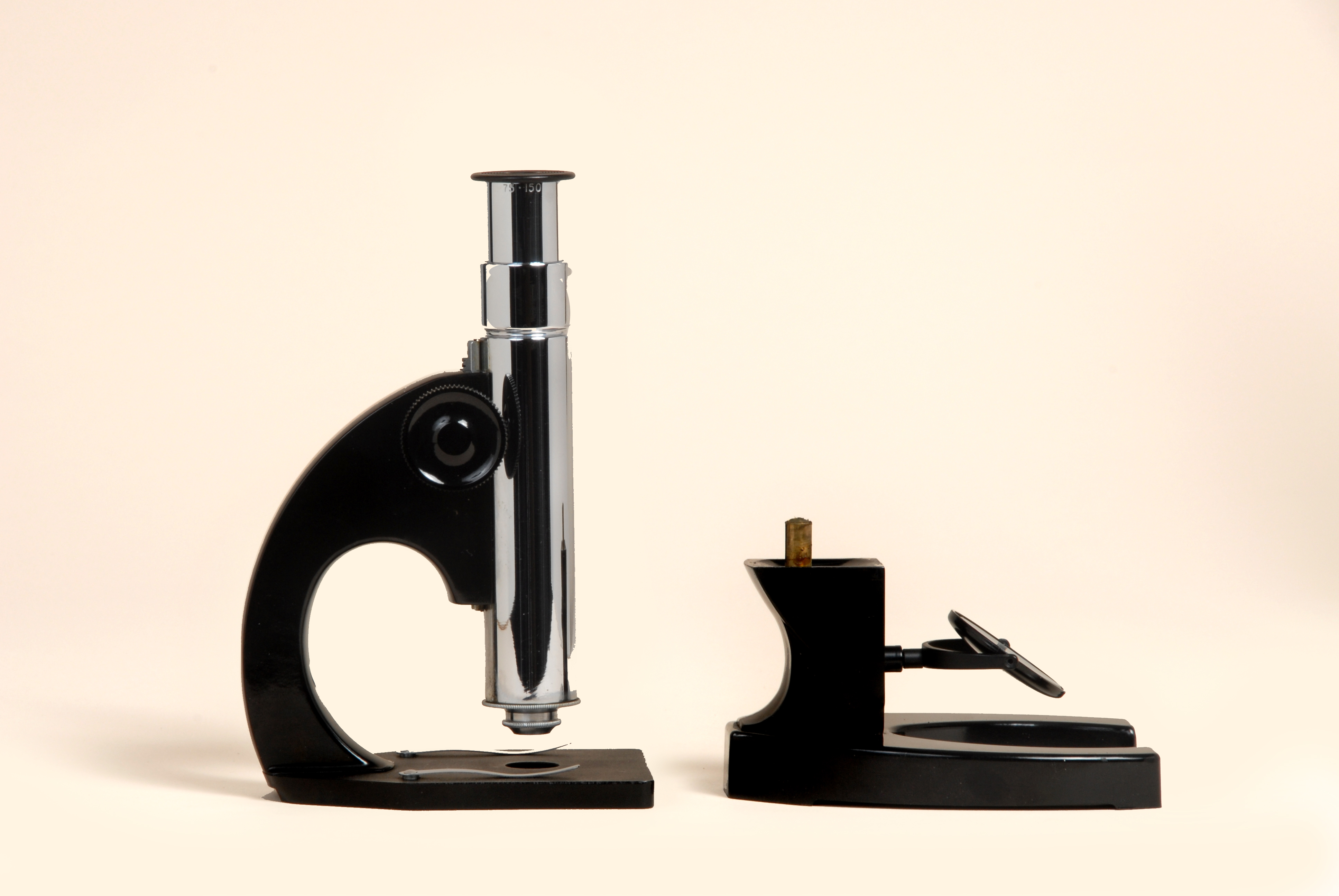
A very interesting feature of this microscope is that the pillar and base separate from the stage and bodytube (Figure 16) in the same way that Bausch & Lomb’s, and other manufacturers’, stereomicroscopes of the time separate from their transmitted-light bases for use with incident light. The substage mirror of the Model R can be removed from the base, and mounted suprastage in a hole provided for it in the limb in order to supply reflected light for illuminating opaque objects, metal surfaces, postage stamps, insects, flowers, etc. (Figure 17). Without its base, the microscope may be placed directly on large objects for examination of their surfaces.
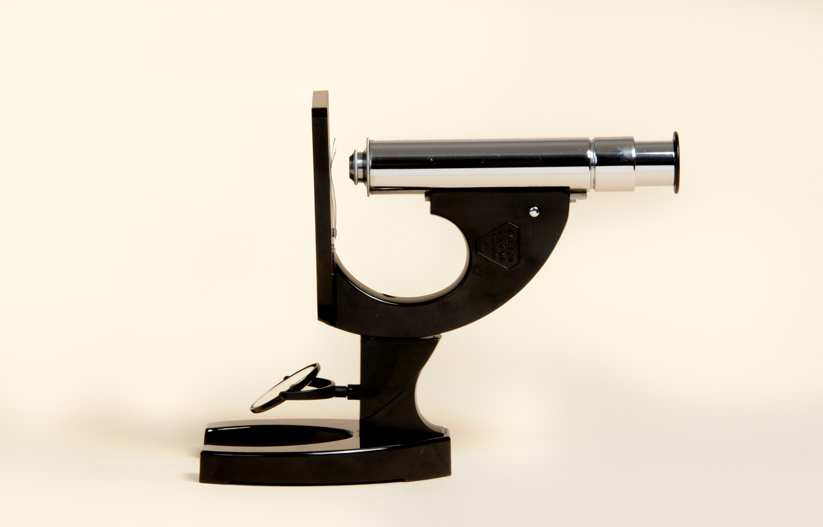
In a clever bit of design engineering, the stage and bodytube, once separated from the base, can be re-attached to the base with the bodytube oriented horizontally (Figure 18). This configuration made it easier to use in conjunction with the box cameras of the day for photomicrography, or for viewing aquatic life in a microaquarium. Additionally, Bausch & Lomb made a separate, optional photomicrography stand for this microscope; in the 1930s, this photomicrography stand, with box camera that used No. 127 film, sold for $12.00.
During the 1930s, Bausch & Lomb also offered another microscope for amateurs, which was designated the Gem. This microscope was of somewhat smaller and simpler construction than the Model R, and, therefore, less expensive. The Gem was available in a kit that was advertised as a 49-piece portable laboratory for mounting specimens, staining, etc. The kit, without microscope, sold for $9.50.
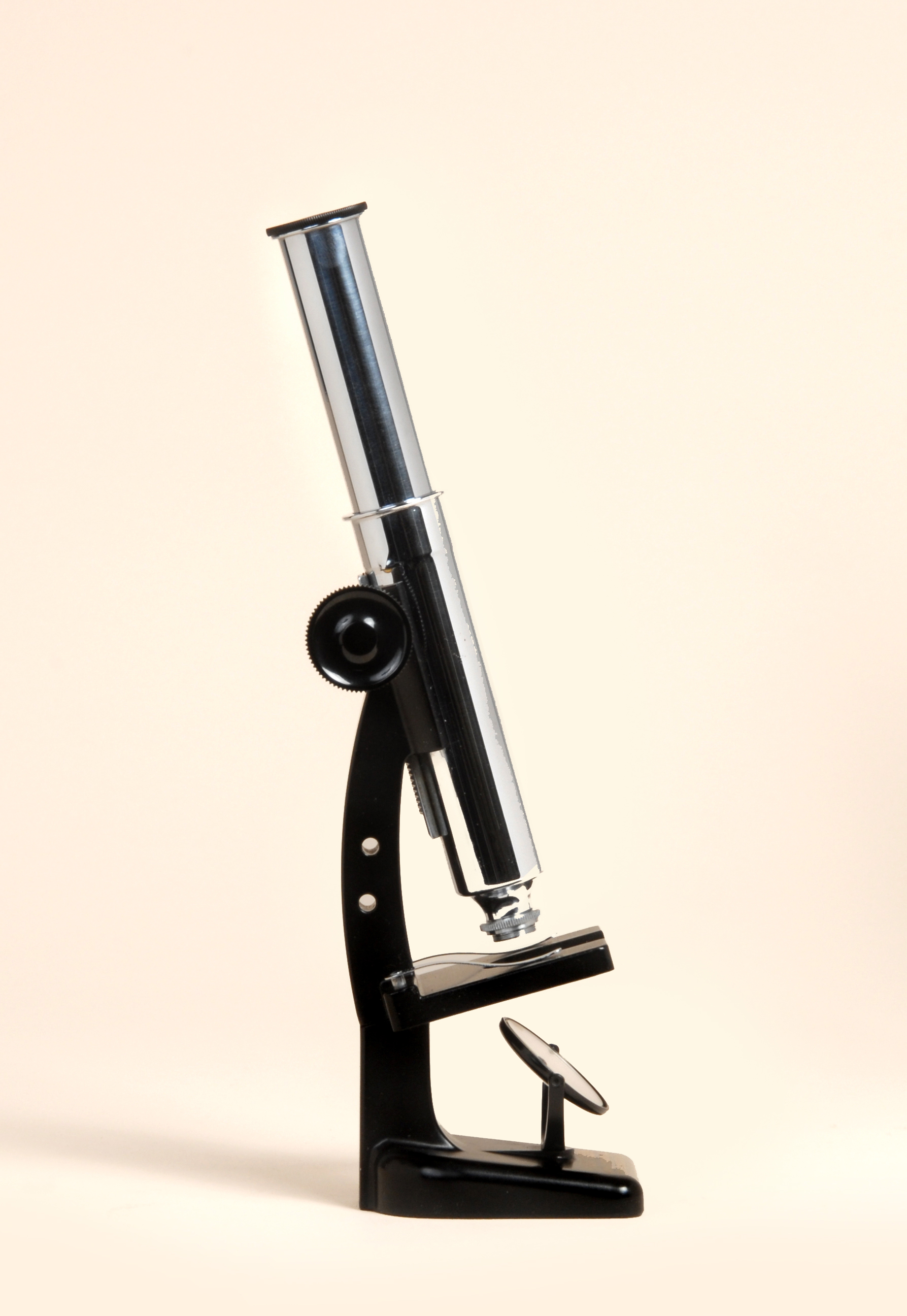
The Gem was quickly followed by the improved New Gem. Bausch & Lomb’s New Gem had its stand cast in inclined position, with its mirror mounted on a vertical support; it was supplied in a handsome walnut box (Figure 19). The New Gem achieved its lowest magnification with the drawtube fully closed, and the lowest magnification objective in place (Figure 20). Maximum magnification was achieved with the drawtube fully extended (Figure 21), and the highest magnification objective installed. Unlike the Model R, the stage and bodytube do not separate from the base, but like the Model R, the mirror can be removed from the base, and remounted above the stage for illumination of opaque specimens (Figure 22).
Available, but rather scarce, was an optional three-part divisible objective (Figure 23), which was capable of multiple magnifications through disassembly and reconfiguration of its three lenses.


Advertisements
Bausch & Lomb’s Model R, New Gem, photomicrograph apparatus, and their Science Kits were extensively advertised during the 1930s. The ads appeared in virtually all of the amateur science hobbyist magazines of the time, including Modern Mechanix, Practical Microscopy, and Popular Science. Indeed, it was the popularity of microscopy among amateurs that no doubt inspired the editorial staff of Popular Science Monthly to compile Wonders Through the Microscope (1934), which was a complete manual for amateurs on how to make their own microscope slide mounts, slide boxes and cases, slide-ringing table, microtome, dissecting microscope, light source, color filter holder, bell jar, warming stand, arc lamp, camera lucida drawing attachment, photomicrographic camera, and microprojector.
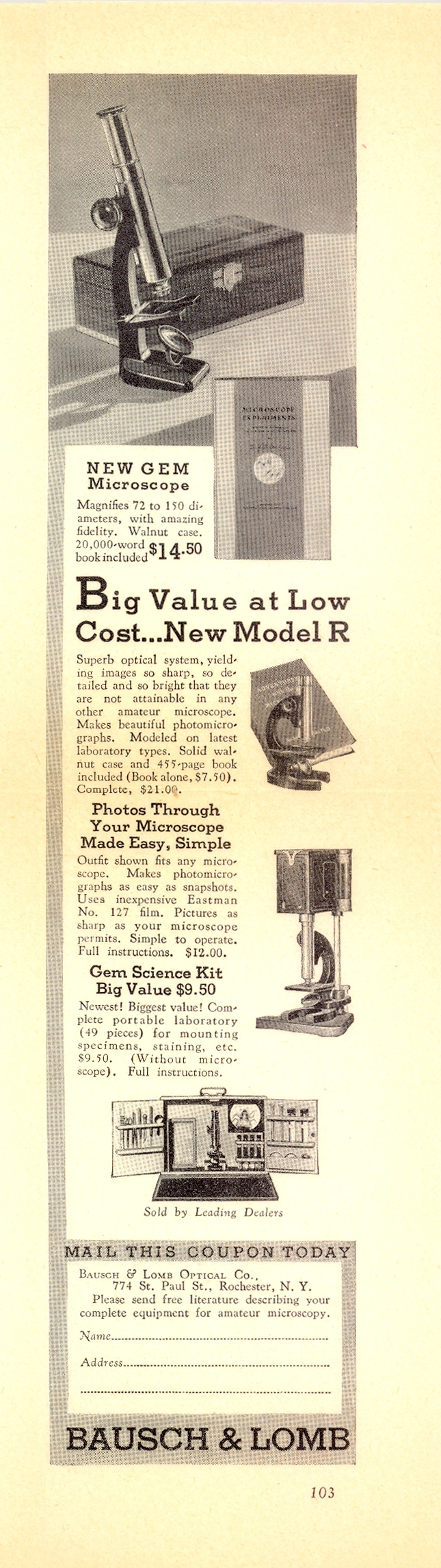
Figure 24 illustrates a typical Bausch & Lomb ad from the late 1930s, offering Corrington’s Adventures with the Microscope ($7.50), the Model R microscope with the book ($21.00), the New Gem microscope with the book ($14.50), the photomicrographic stand and camera ($12.00), and the Gem Science Kit without microscope ($9.50).
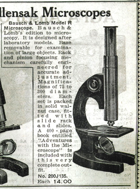
A 1939 ad is shown in Figure 25, with details on the Bausch & Lomb Gem microscope (Figure 26), and on the Model R microscope (Figure 27). The Wollensak microscope also shown in this ad is from Bausch & Lomb’s
down-the-street-in-Rochester competitor.


For additional ads and observations on these amateur microscopes, see Charles Gellis’ fine article in the Spring 1998 issue of the Journal of the Microscope Historical Society (Gellis 1998). Manuel del Cerro has also written about the Bausch & Lomb Model R microscope, and includes a copy of the Directions for Use…; his article, which also contains some information about the Wollensak microscope, is in the Winter 2000/2001 issue of the Journal of the Microscope Historical Society (del Cerro 2000/2001).
Conclusion
Julian D. Corrington’s contributions to amateur microscopy are today just as valuable and inspiring as they were when he first offered them to the public. It was fortuitous that the New York Microscopical Society obtained Corrington’s autobiographical note in 1978, the same year in which Corrington’s microscopical contributions were recognized by the State Microscopical Society of Illinois in naming him the recipient of their Honorary Award, for Corrington passed away in Miami-Dade County, Florida in November 1979.
Julian D. Corrington was a professor of zoology at the University of Miami, and served as chairman of the zoology department. He taught zoology and botany from 1926 to 1967, and also taught the university’s first courses in marine zoology and marine botany during the 1930s. The University of Miami maintains the Julian D. Corrington papers, 1917-1963. The 17 boxes of class records and course materials, dated 1944-1963, include syllabi, memos, lecture notes, correspondence, etc. are available for research.
Selected References
Corrington, Julian D. (1922) The Winter Birds of the Biloxi, Mississippi Region. The
Auk 39 530-556.
Corrington, Julian D. (1927) Commensal Association of a Spider Crab and a Medusa. Biological Bulletin 53 346-350.
Corrington, Julian D. (1930) Morphology of the Anterior Arteries of Sharks. Acta Zoologica 11 (2-3) 185-261.
Corrington, Julian D. (1931) Abnormal Herpetological Specimens from Syracuse, New York. American Naturalist, 65 (696) 77-85.
Corrington, Julian D. (1934) Adventures With the Microscope. Bausch & Lomb Optical Company, Rochester, New York, 455 p. 350 figures. Photos by Charles S. Foster, Eastman Kodak Company.
Corrington, Julian D. (1939) The Microscope, A Pictorial History. Nature Magazine, February 1939. Reprinted from a copy supplied by Charles Gellis in Journal of the Microscope Historical Society7 (2) Summer 1999, 29-38.
Corrington, Julian D. (1941) Working with the Microscope. Whittlesey House, McGraw-Hill, New York and London, 418 p. At least seven printings.
Corrington, Julian D. (1957) Exploring With Your Microscope. McGraw-Hill, New York, London, Toronto, 229 p. At least five printings.
Corrington, Julian D. (1959) Experiments Using the 3-D Microscope. Bausch & Lomb Optical Company, Rochester, New York, 11 p.
Corrington, Julian D. (1965) “New Worlds of Entertainment” on the Microscope Stage; Experiments performed and recorded with Tasco Educational Kits. Tasco Sales, Inc., Miami, Florida, 64 p.
Corrington, Julian D. (1969) Getting Acquainted With the Microscope. Bausch & Lomb, Inc., Rochester, New York, 47 p.
del Cerro, Manuel. (2000/2001) The Bausch & Lomb R Microscope, A Youngster’s Dream With a Note on its Competitor From Around the Block, The Wollensack (sic) 425 Power and Others. Journal of the Microscope Historical Society 8 (3) Winter 2000/2001 83-103. Includes a copy of the Directions for Use of
the Bausch & Lomb Model R Microscope, Cat. No. 81-72-03-01 (1938).
Delly, John Gustav. (1999) Julian D. Corrington and the Bausch & Lomb Model R Microscope. State Microscopical Society of Illinois: μ • Notes July 1999 15-20.
Editorial Staff of Popular Science Monthly (1934) Wonders Through the Microscope; A Complete Manual for Amateurs. How to use equipment, secure and preserve specimens, take photomicrographs, etc. Popular Science Publishing Company, New York, 192 p. First edition February 1934; Second edition August 1934; Third edition May 1938.
Gellis, Charles. (1998) In Pursuit of the Elusive Model R Microscope; Observations and Measurements: The Technical Side of the New Gem and the Model R. Journal of the Microscope Historical Society 6 (1) Spring 1998, 12-22.
Nature Magazine; An Illustrated Monthly. Vols. 1-52, 1923-1959. Corrington wrote and edited the column “Under the Microscope” for this magazine for twenty-two years.
Santacana, Guido E. (2000) A Complete Student Microscope Set from 1965. 4 p. Micscape
Magazine. See, also, Santacana’s comments at: http://groups.yahhoo.com/group/Amateur_Microscopy/message/1715 accessed 16 September 2012.
Wren, L.A. and Corrington, Julian D. (1963) Understanding and Using the Phase Microscope; including “Experiments With the Phase Microscope.” Unitron Instrument Company, Newton Highlands, Massachusetts, 59 p + 5 p. ads. 2 sections, 7 chapters. Experiments by Corrington begin on p. 46.
Comments
Walter Renn
Dear McCrone Group,
Thank you for this generous biographical note on Dr. Julian Corrington. He was one of my two most favorite undergraduate professors. I took him for Comparative Anatomy and Embryology and gained my
first understanding of evolutionary theory in those classes. He was very memorable and influenced my professional life strongly. I went on to gain my Ph.D. and am recently retired in Florida. He remains my hero!
add comment