Microscope Activities, 23: Ultraviolet Fluorescence Microscopy
In the past, Hooke College of Applied Sciences offered a microscopy workshop for middle school and high school science teachers. We thought that these basic microscope techniques would be of interest not only for science teachers, but also for homeschoolers and amateur microscopists. The activities were originally designed for a Boreal/Motic monocular microscope, but the Discussion and Task sections are transferable to most microscopes. You may complete these 36 activities in consecutive order as presented in the original classroom workshop, or skip around to those you find interesting or helpful. We hope you will find these online microscope activities valuable.
EXPERIMENT 23: Ultraviolet Fluorescence Microscopy
Goal
To assemble an inexpensive source of ultraviolet light, and make observations of fluorescence.
Level
Basic-Intermediate
Materials Needed
60W Black Light Bulb or Mini Fluorescent Lantern
Procedure
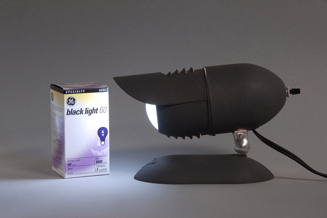
Obtain a 60-watt black light bulb, and install it in place of the regular white 60-watt bulb in a gooseneck desk lamp, or in a microscope illuminator that is equipped with an Edison screw base. The Bausch & Lomb microscope illuminator illustrated in Figure 23-1 was purchased online for $12, and the regular 60W bulb that was installed was replaced with a 60W black light purchased at ACE Hardware for under $5.

This modified illuminator can now be used with traditional microscopes that are equipped with a mirror to illuminate any sample with ultraviolet light.
For use with the Boreal/Motic microscope, a 1” round or square mirror from a craft shop will have to be placed above the light exit port to reflect the ultraviolet light up into the condenser (swing the lower ground glass out of the way), or the UV light from the lamp can be directed downward from above the stage for epi-fluorescence.
Another inexpensive source of ultraviolet light in a Mini Fluorescent Lantern sold in fishing supply stores or in the fishing section of large department stores (Figure 23-2).

This kind of lamp or lantern that uses a mini fluorescent tube coated for ultraviolet light output is used by fishermen for night fishing; the fishing line has a fluorescent compound introduced in manufacture so that the location of the line can be spotted at night if illuminated with ultraviolet light. Again, the ultraviolet light can be directed up into the substage condenser via a mirror, or held above stage for epi-fluorescence.
Figure 23-3 shows the ultraviolet light lantern being handheld above samples of fluorescent minerals. This same kind of handheld UV illumination works especially well with stereomicroscopes and larger samples at lower magnifications.

Discussion
Ultraviolet fluorescence is an extremely important and useful technique in analytical microscopy and biomedical research. Professional fluorescence microscopes utilize high pressure mercury vapor lamps as their source of ultraviolet. However, as a safety note, it is imperative to realize that the mercury vapor lamp gives off wavelengths in the short ultraviolet, ~254 nm, which have germicidal action, and are harmful to the user’s eyes. Normally, excitation filters are placed in the light path that isolates only the Long Ultraviolet, ~365 nm, and even then barrier filters are installed somewhere above the fluorescing sample, but before the eyes.
The inexpensive sources suggested here are wideband Longwave ultraviolet light centered around the 365 nm region, with considerable violet from the visible region. They are, nevertheless, a very useful and inexpensive introduction to the use of ultraviolet light in fluorescence microscopy.
In recent years, advances in light-emitting diode (LED) technology have extended the wavelength range down into the ultraviolet. UV-LEDs are now quite common; indeed, today there are available arrays of UV-LEDs made into light sources for transmitted ultraviolet microscopy. These UV-LED sources do away with the need for high-pressure mercury-vapor arc lamps. This new form of ultraviolet illumination can be experienced through the use of an inexpensive blacklight keychain flashlight that utilizes a UV-LED, and is powered by four tiny AG3 (or AG 13) batteries; the peak wavelength is 395 nm.
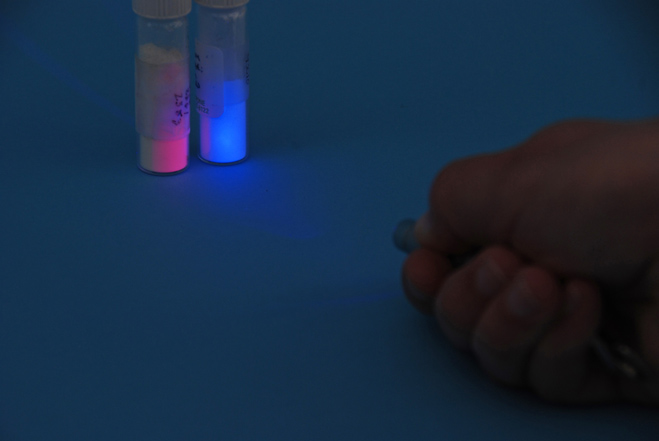
Figure 23-4 illustrates one such UV-A light source that is only 4.5 cm long [Crazy Aaron’s Putty World]; there is a push-button in the side of the body that turns the unit ON.
Figure 23-5 shows the UV-A light source in use, activating two vials of phosphors. For use with the compound microscope or the stereomicroscope, the light source is merely held by hand close to the specimen, directing the UV light toward the sample.


Task
Purchase or make a source of longwave ultraviolet light, and examine a number of substances with it. Be sure to look at chlorophyll-bearing algae from the aquarium, and report the UV fluorescence color of chlorophyll. What else in the aquarium sample fluoresces? Note that you can introduce fluorescein into your sample drop, or other “fluorochromes” so as to induce fluorescence where it does not occur naturally.
Some things fluoresce in and of themselves naturally; this is referred to as “primary fluorescence” or “auto fluorescence,” when fluorochromes are added to induce fluorescence, it is referred to as “secondary fluorescence” or “induced fluorescence.”
You will notice that Canada Balsam and a number of other mounting media fluoresce, making permanent slide preparations difficult to evaluate because of the masking effect of overall fluorescence.
For dust particle characterization and identification, samples are mounted in water, glycerin, or “fluorescence-free” or “low fluorescence” immersion oil.
Comments
add comment