Nitzschia reversa Photographed Through A Student Microscope
The story of reworking a Monolux microscope into a universal student microscope with fiberoptic source illumination is covered in my first article on microscopy (1). I am now preparing for my daughter and granddaughter to inherit this microscope. My microscopy studies in the past 14 years have been with my modified LOMO Biolam. I know my family’s interest is in looking at water microorganisms. I decided that the modified Monolux with its Edmund objectives will be easiest for them to use initially. The Biolam with the LOMO objectives equipped with water immersion caps and the water sample contained in a microaquarium slide can be used once enough experience is gained with the Monolux (2). The early use of the Monolux was with darkfield stops for the 40X 0.65 NA and 60X 0.85 NA objectives mounted in caps fitted over the aperture diaphragm on the output end of the fiberoptic light guide. More recently, a slider with stops was incorporated into the illumination system, as shown in Figure 1.
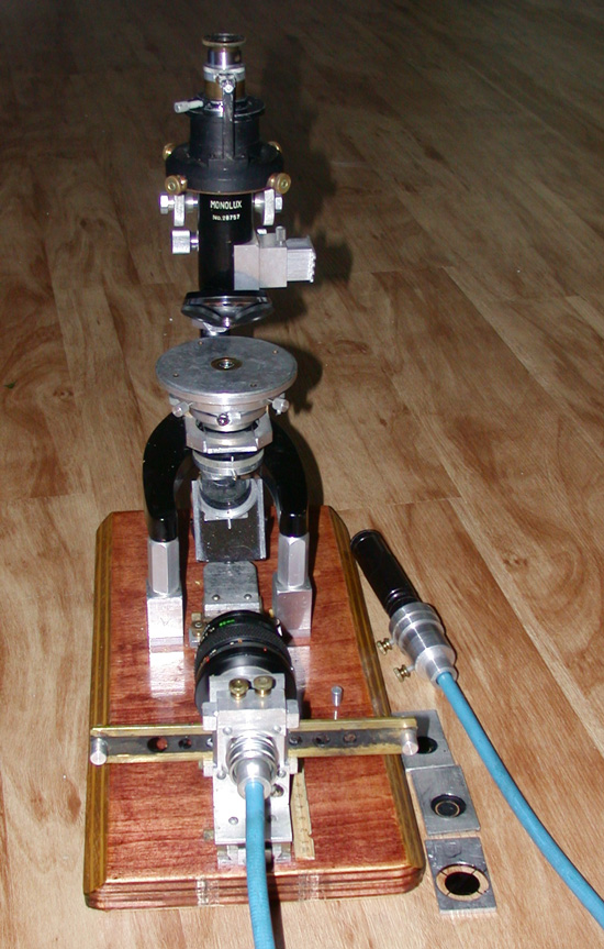
Also, additional stops for the filter slot of the condenser were made for the 40X and 60X objectives. The rotating stage made for the Monolux facilitates use of immersion oil or glycerin to couple the slide to the top of the condenser. The close fitting brass cap through the center of the rotating stage keeps the oil or glycerin from seeping down the side of the condenser. The input end of the fiberoptic light guide can now be coupled with either a 150 watt quartz halogen illuminator or an LED flashlight. The condenser has very significant spherical aberration at higher illumination NA as demonstrated in one of my earlier articles (3). This deficiency precludes Köhler illumination for quality brightfield imaging with high NA objectives. This article on a multimode condenser for the Biolam demonstrated high resolution, high contrast imaging for an uncorrected Abbe condenser using stops for darkfield and circular oblique lighting (COL).
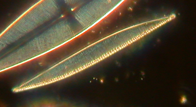
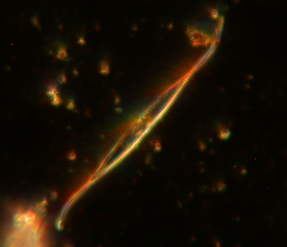
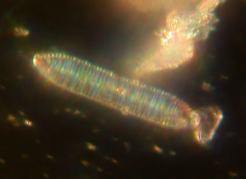
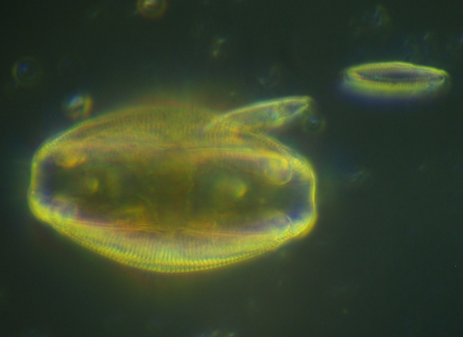
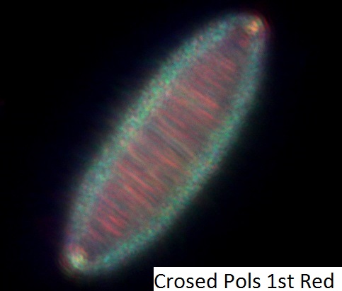
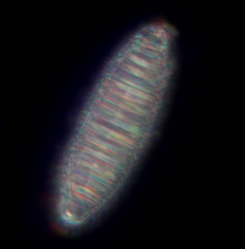
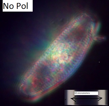
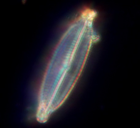
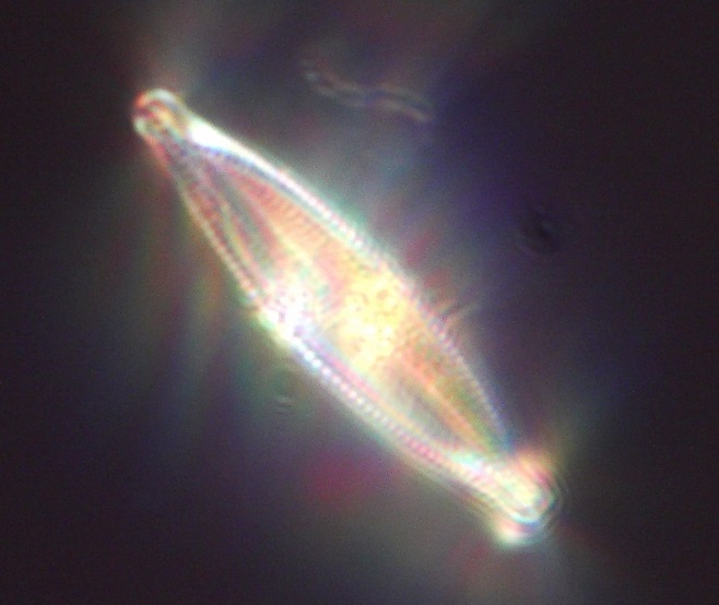
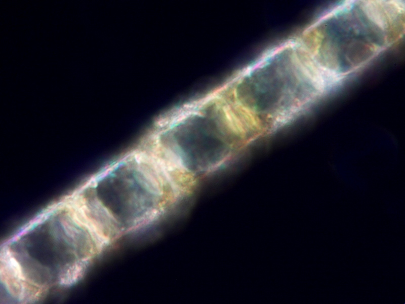
I was curious about the high resolution capability of the Edmund 60X 0.85 NA in COL and darkfield. My test specimen was the Klaus Kemp 8-Form Diatom Test Slide. The theoretical resolution of a 0.85 NA objective, with the zero and first order diffraction rays falling on opposite sides of the outer edge of the back focal plane of the objective, is about 0.33 micrometers. The reported striae spacing for the diatom Nitzschia sigma is 0.43 micrometers. The puncta spacing along the striae is significantly less than the striae spacing because the stria can be just resolved with a 0.65 NA objective using COL (4). The puncta of Nitzschia sigma were resolved with the 0.85 NA objective and COL as shown in Figure 2. The properly-sized stop had to be inserted into the condenser filter slot. The tube length marked on the objective is 170 mm, but best image sharpness was obtained with a 160 mm tube length used for the resolution tests. The resolution in darkfield is somewhat less, see Figure 3. Best darkfield contrast required annular illumination with a stop at the end of the light-guide source, where the aperture diaphragm is located, as well as a stop in the condenser slot. These resolution tests were conducted after a convex deposit of glycerin was applied covering the shallow depression on the top lens of the condenser and covering the top of the brass cap. The slide was then applied over the glycerin; air bubbles were not a problem.
Click on any image to view a gallery of larger images.

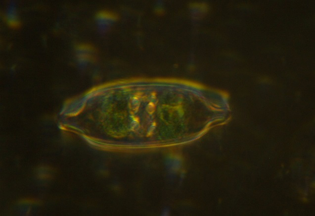
Darkfield with the 60X Edmund objective was subsequently used to look for interesting diatoms and desmids in water samples. The best source of diatoms of many different types was found to be water sucked from near the algae covered rocks along the shore of the Cache La Poudre river on the eastern edge of the Colorado city of Fort Collins; this is unpolluted fresh water. Interesting diatoms are photographed to share with family and friends. A disposable pipette is used to suck up specimens for microscopy from the bottom of the jar of river water. A 2” X 3” slide is coupled to the top of the condenser with glycerin and a drop of specimen is deposited over the condenser top and covered with a 22 mm X 40 mm coverglass. Traces of glycerin get on my fingers, but is much more easily removed than immersion oil.
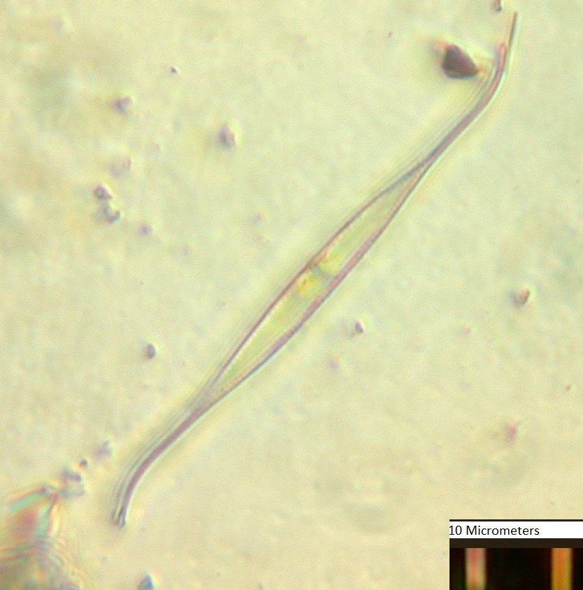
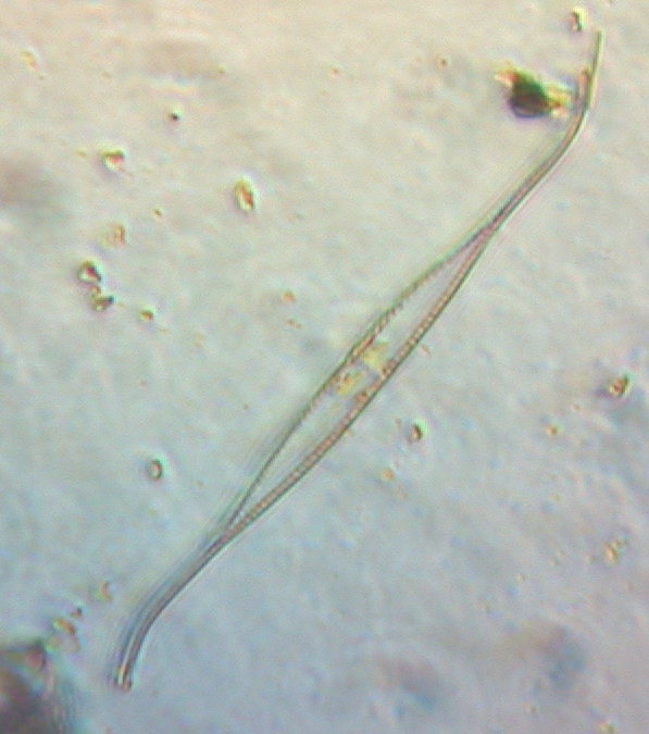
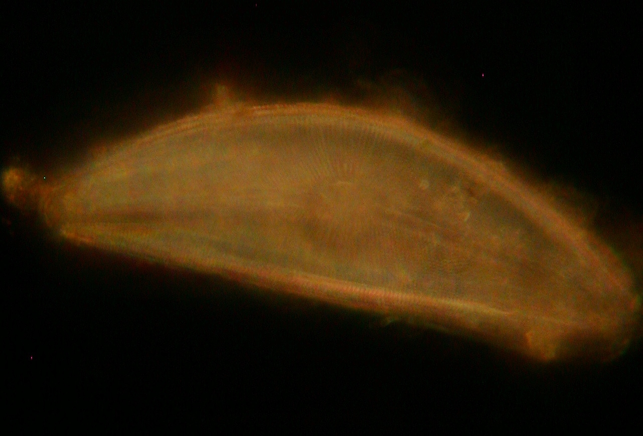
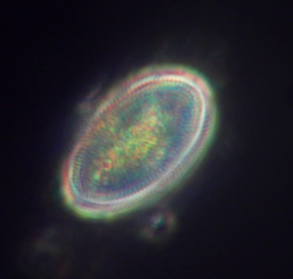
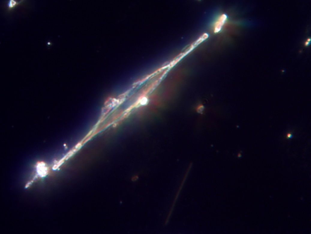
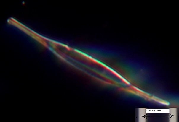
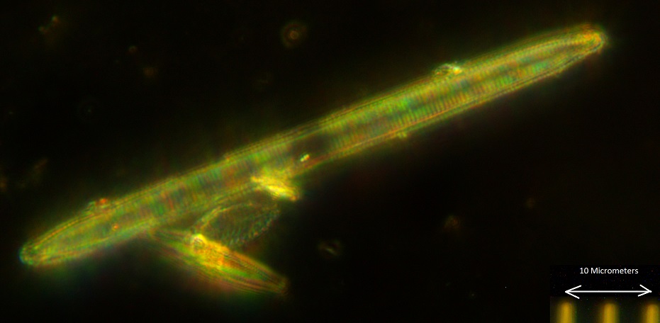
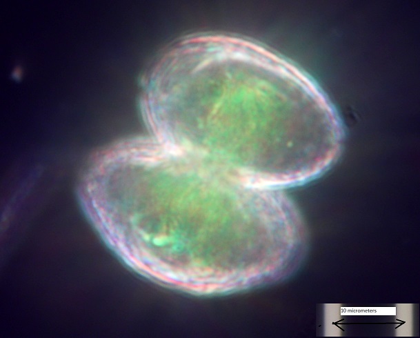
An unusually shaped diatom is common in the river water. The slender diatom has reverse hook ends as shown in Figure 4. Figure 5 shows the same field with COL illumination. David Walker, editor of Micscape web microscopy magazine, helped to identify it as Nitzschia closterium. The problem with this identification is that Nitzschia closterium is found in sea or brackish water. The W. Smith classification for Nitzschia reversa seems to be a better fit and is listed as being a freshwater diatom, but not accepted by the diatomist community.
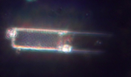
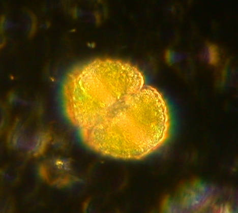
Enjoy these additional darkfield images. Click on any image to view a gallery of larger images.
Additional images were provided by the author and published at his request. Click on any image to view a gallery of larger images.


References
- Clarke, T. M. (2000) Building an Affordable Universal Student Microscope.
The Microscope, 48:1 19-38.
- Clarke, T. M. (2000) Water Immersion Caps for Pond Water Microscopy.
The Microscope, 48:2 87-91.
Comments
add comment