JEOL NeoScope JCM-7000
SKU:NEOSCOPE-7000
Are you considering purchasing a JCM-7000 benchtop SEM? Contact us for your RFP or quote.
Increase your scope with the JCM-7000 benchtop SEM. The perfect complement to light microscopy, this compact, tabletop SEM with a magnification range of 10X – 100,000X gives you the power of scanning electron microscopy in a convenient package.
Equipped with a large sample chamber, both high and low vacuum modes of operation, both secondary and backscatter electron detectors, real-time 3D imaging, easy to use metrology tools, and optional fully-integrated energy dispersive X-ray (EDS), the JCM-7000 NeoScope is smart, flexible, and powerful.
- Magnification:
- 10X – 100,000X (print 128 mm x 96 mm)
- 24X – 202,168X (display 280mm x 210mm)
- Minimize sample prep and generate images in less than 3 minutes
- Select automatic conditions, or set and save your own
- Optional EDS for elemental analysis
New — Zeromag

- Zeromag simplifies navigation and enhances throughput
- With Zeromag, you can navigate from a color optical image—as you increase the magnification, you transition from the optical to live SEM image automatically
- Set up large area automated image montage and stitching; EDS option includes automated montage X-ray map
- Automatically link SEM image, position, optical image, and EDS data (with EDS option)
New — Live 3D Imaging

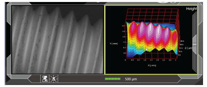
With the EDS option, our 6-channel, high sensitivity, solid state backscatter electron detector acquires composition, topographic and shadow (combination of composition and topography) images, and supports live 3D imaging.
Combine the live 3D image with software to create a 3D model and calculate surface texture data, including:
- Cross-sectional profile
- Height
- Surface roughness
New — Live Elemental Analysis
When you opt for the EDS option, our analytical model includes JEOL’s fully-embedded EDS system with real-time EDS spectra during image observation. With live elemental analysis, you can:
- View EDS spectra in real time as you search for an area of interest
- Set analysis points, area, map positions, and line scans
- View major elements detected, and automatically display on live EDS window
For over 50 years, McCrone Microscopes & Accessories has remained the industry expert. Let us power up your benchtop to give you remarkable images of protein structures, nano particles and stress fractures.
Contact us for a RFP or quote for your benchtop SEM.
What is a tabletop SEM / benchtop SEM?
In brief, a tabletop SEM is a portable, benchtop scanning electron microscope used for on-site analysis, including materials science and biological samples. Like all scanning electron microscopes, a tabletop SEM uses a narrow, focused electron beam to scan a sample’s surface, generating a 3D image. As electrons interact with atoms within the sample, information on the sample’s topography and composition are produced.
- Tabletop SEMs are portable for on-site analysis
- Narrow, focused electron beam generates a 3D, high depth-of-field image highlighting a sample’s surface structure
- The signal from low-energy secondary electrons (SE) produces a detailed topographical image
- Backscattered electrons (BSE) reveal information about the distribution of elements within a sample
5 Benefits of the JEOL-7000 Tabletop SEM vs. a Full-sized Instrument:
- A benchtop instrument has advanced electron optics technology with a nearly equivalent performance to a full size SEM, at a lower cost.
- Its convenient, small size allows for a portable instrument with no special environmental requirements—optimal for quality control applications.
- A tabletop SEM produces quality, high resolution images without needing a large footprint.
- The JEOL JCM-7000 is a turnkey solution for gathering information about unknowns, including real-time elemental analysis.
- You can bring the instrument to the sample—perfect for on-site analysis in academic and educational settings.
Options Offer More Robust Performance
- An EDS detector option is available for elemental microanalysis
- Addition of our color optical camera (stage navigation system) option provides seamless sample navigation from the optical to high resolution SEM image
- Supports real time 3D imaging, and combined with optional software, various surface texture data can be calculated such as: cross sectional profile, height, surface area, surface roughness and more
- In addition to the popular tilt/rotation motorized holder (T =-10° to 45°, R = 360°), custom and specialty holders are available
- An oil-free diaphragm pump (replaces rotary pump)
Features
- Magnification – 10X – 100,000X (print 128 mm × 96 MM)
- Observation modes – High vacuum/low vacuum
- Electron gun – Tungsten filament; Wehnelt integrated
- Landing voltage – Selected from software interface: 15 kV/10 kV/5 kV
- Specimen stage – X-Y manual control; X: 35 mm; Y: 35 mm
- Maximum specimen size – Diameter 80 mm, height 50 mm
- Detectors — SE: Everhart-Thornley; BSE: 6-channel solid state
- Signal detection – High vacuum (secondary electron image backscattered electron image); low vacuum (backscattered electron image)
- File format – TIFF, BMP, JPEG, PNG; AVI for live image display
- Image shift – Electromagnetic; approx. ±50 µm in XY (WD 12 mm)
- System OS – Windows®10; Intel®Core™ i5 CPU (or equal)
- Automated functions – Auto focus, auto stigmator, auto contrast/brightness control, auto gun alignment
- Operation – Touch panel, mouse/keyboard control of GUI
- Optional accessories – Tilt/rotation motor drive, stage navigation system, 3D analysis software, off line data analysis software, energy dispersive X-ray spectrometer (EDS) with 30 mm2 silicon drift detector, EDS particle analysis software, oil-free diaphragm pump (replaces rotary pump); custom and specialty holders available
- Base unit – 12.8″ (W) x 23.1″ (D) x 22.3″ (H) (in mm: 324 x 586 x 566)
- Power supply – Single phase AC 100 V (compatible with 120 V, 220 V, 240 V); fluctuation ±10% or less, with grounding
These webinars feature the JEOL JCM-7000. Enjoy!
Meet the JEOL JCM-7000 NeoScope Benchtop SEM
Which Microscope Should I Use? A Multi-tiered Microscopy Analysis on Bandages/Wound Care Products
Read “Using Benchtop Scanning Electron Microscopy as a Valuable Imaging Tool in Various Applications” in Microscopy Today, September 2022.
Speak to a Scientist
McCrone Group's analytical scientists are available to answer your questions. Have a project you'd like to discuss? Give us a call or email us!
