Simon Henry Gage: Quintessential American Microscopist
[This article is based on one that first appeared in The Journal of the Microscope Historical Society, Volume 17, 2009, and is reprinted here by kind permission of that journal’s editor, Daniel Kile]
Introduction
Simon Henry Gage (1851-1944) was one of the most remarkable, influential, and important figures in the history of American microscopy. Every serious user of the microscope will, sooner or later, become familiar with him either through one of the approximately 200 publications in his bibliography, or, more likely, from possession of one or more of the seventeen editions of his book, The Microscope, that was continuously available for some 60 years.
A Brief Biography of Simon Henry Gage
Simon Henry Gage was born on May 20, 1851 in Milford Township, Otsego County, New York. At the time, this was a forested area surrounding Lake Crumhorn. The family moved from this pioneering area to the village of Worcester in order to be nearer to schools. Young Gage attended Charlottesville Seminary and the State Normal School at Albany. To obtain a financial start for college he worked as an itinerant tent-photographer, in the process learning photographic skills that he would later use in research and teaching.
Gage entered Cornell University in 1873, originally planning on becoming a physician. To help support himself, he became student assistant to Dr. Burt Green Wilder, who was Head of the Anatomical Department, which then included the subjects of physiology and hygiene, zoology, and comparative anatomy; he continued in this position throughout his undergraduate years. While still an undergraduate, Gage began teaching other students in the newly introduced biology courses. In the Autumn of his graduation year he became an instructor at Cornell, having full charge of the work in the Department of Comparative Anatomy and Microscopy. He graduated in 1877 with a B. S. degree in Natural History. His graduation thesis, which was on the life history of the Cayuga Lake Stargazer (Cottus), became his second published paper. Gage was active in the American Microscopical Society, which grew out of the first National Microscopical Congress, convened in 1878; he would later twice serve as the Society’s President.
By 1881 Gage had become Assistant Professor of Physiology and Lecturer in Microscopical Technology at Cornell, and in December of that year he married Susanna Stuart Phelps, one of his students who had graduated the year before (she helped him in his work, doing everything from pen drawings to making enlarged three-dimensional models of serial sections on her sewing machine). Their son, Henry Phelps Gage, was born in October 1886. Susanna died in 1915.
Textbooks were scarce, so Gage helped Dr. Wilder write a book on cat anatomy, and started his Outline of Microscopical Technology—a hectographed laboratory guide for his students that would evolve into his famous textbook. The fashion at that time was to go abroad for advanced work, but Gage was not financially able to do this, so he attended all the meetings held by anatomists, zoologists, and the American Association for the Advancement of Science; he felt that this gave him the best and broadest kind of post-graduate training. In 1885 Gage was made Chairman of the section on Microscopy and Histology of the American Association for the Advancement of Science. During a sabbatical year in 1889 Gage finally got his chance to study at Göttingen and at the University of Berlin; he also visited Paris and London.
Gage lived through a time of great changes in the development of specialty subjects. In the 1860’s, when the modern medical schools started in Europe, especially in Berlin, Vienna, and Paris, there were only six or seven study subject areas—there would be at least fifty a hundred years later. These changes were reflected in Gage’s many academic titles and teaching subject areas over the years as “natural history” expanded into botany, zoology, comparative anatomy, histology, embryology, entomology, etc. From 1889 to 1893 Gage was Associate Professor of Physiology and Lecturer on Microscopical Technology at Cornell; from 1893 to 1895 he was Associate Professor of Anatomy, Histology, and Embryology.
Many readers will be familiar with the natural history books published by The Comstock Publishing Company. This company was founded by Professor John Henry Comstock and Professor Gage in 1893 to make reasonably priced books on microscopy, histology, and entomology available to students. Through the bequest of Comstock and the gift of Gage this publishing company became the property of Cornell University in 1931.
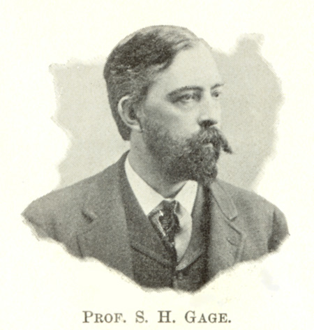
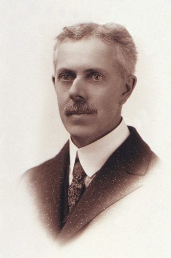
In 1895, Gage was elected President of the American Microscopical Society. In the following year he was made President of the New York State Teachers’ Association. The Journal of Applied Microscopy for September 1898 (J. Applied Microscopy 1898) mentions in its report on the 21st annual meeting of the American Microscopical Society that Gage was a Member of the Executive Committee (1898-1899), and that he presented a paper on Some Laboratory Apparatus for Histology; this notice is accompanied by a portrait of Gage (Figure 1), who would, at this time, be 47 years of age.
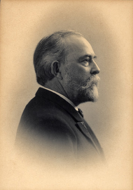
When the new Veterinary College building was completed in 1896, Gage moved into it, and had a lecture room equipped with microprojection apparatus—a topic he and his son would later write a book about. In addition to his teaching and research, Gage was instrumental in establishing laboratories and libraries. The new Veterinary College was in need of a library, and Gage was able to obtain funds from the widow of the then late Governor Roswell P. Flower, and added to the Governor’s own contribution toward establishing the Flower Library in 1899. In that Library today there hangs a magnificent oil portrait of Gage painted in 1920 when Gage was 69 years of age (Figure 2, Courtesy of Cornell’s College of Veterinary Medicine Archives/Image Lab).
At the turn of the century, Gage was involved in the founding and editing of the American Journal of Anatomy. In 1906 he was elected President of the American Microscopical Society for the second time. His presidential address, Origin and Development of the Projection Microscope, would not only indicate his interest in history, but would presage his book on microprojection.
Gage retired in 1908 as Professor Emeritus of Histology and Embryology, after 25 years of teaching at Cornell, intending to devote the remainder of his life to research. One of his first tasks was to publish the microprojection methods he had used in his teaching; he wrote this with his son Henry; Optic Projection was published in 1914 (GAGE and GAGE 1914). Gage then started a history of optics, adding a chapter to the 12th edition of The Microscope (GAGE 1917).
In 1918 Gage, who had already been retired for ten years, returned to teaching to relieve the shortage of teachers during the World War I years. At the 1920 meeting of Anatomists, Gage gave a report on his studies of fat digestion—a study he instituted in 1917 on the assimilation of fats, using Sudan III, a fat-soluble dye. Active throughout the 1920’s, Gage was honored on his 80th birthday in 1931, and immediately started his researches on the comparative anatomy of the pancreas (a monograph was finished, but did not appear until after his death).
In 1933 Gage married Clara Covert Starrett; she helped him with the revisions of the last two editions of his book, The Microscope. In 1941, at the age of 90, the 17th revised edition of The Microscope was published—this, 33 years after he retired!
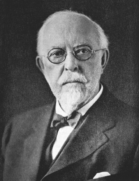
In 1943 Gage started work on his History of American Microscopy; it was almost finished, when on October 20, 1944, at the age of 93, Gage died. Oscar W. Richards edited and published Gage’s Microscopy in America (1830-1945) in 1964 as a supplement to the Transactions of the American Microscopical Society (RICHARDS 1964). As the Frontispiece to this Supplement, there is another portrait of Gage later in life (Figure 3).
This brief biography is based, in large part, on a more comprehensive one written by Oscar Richards that accompanied Gage’s Microscopy in America (1830-1945), which, in turn, was based on a long biography written by Gage’s son, Henry Phelps Gage. Readers desiring to know more of the many facets of Gage’s life are encouraged to read Richards’ account.
The Works of Simon Henry Gage
A detailed account of all of Gage’s publications cannot be made here, as he had 200 published articles, in addition to the seventeen editions of his book, The Microscope, and the book on optic projection written with his son Henry. However, two of the articles will be discussed—one macro and one micro—and all editions of his books.
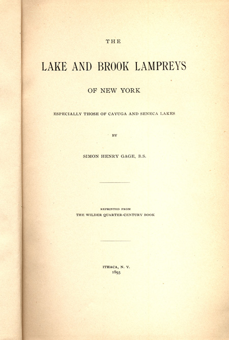
Gage’s early articles are devoted to zoological subjects found in local woods and lakes, as already indicated by his second published paper on the life history of the Stargazer (Cottus)—a kind of mottled sculpin—found in nearby Cayuga Lake. Another monograph-length publication was one on The Lake and Brook Lampreys of New York; Especially Those of Cayuga and Seneca Lakes (Figure 4) reprinted in 1893 from the Wilder Quarter-Century Book (GAGE 1893). Dr. Wilder had initiated these studies with his students, and Gage tells us that he had personally studied lampreys at all stages of life since 1875; i.e., while still a student. All of the local lakes around Cornell have Lake Ontario as their final destination, through the Oswego River, and then through the St. Lawrence River to the Atlantic Ocean. The lampreys, spending their adult life in the ocean, became accustomed to remain permanently in fresh water. I chose this monograph as one to mention because a similar situation persists here in Lake Michigan, and because the lamprey was one of my own subjects of study in Comparative Vertebrate Anatomy in 1951. Gage discusses the life history and anatomy of the lamprey, but the wonderful plates illustrate only the sperm and blood cells from the microscopical standpoint.
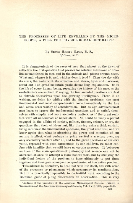
An exclusively microscopical article is one with the intriguing title, The Processes of Life Revealed by the Microscope; A Plea for Physiological Histology (GAGE 1896a) (Figure 5), which was actually Gage’s Presidential Address before the American Microscopical Society. I chose this particular article, again, for personal reasons; having studied Vertebrate Histology, I wondered what Gage meant by “the processes of life revealed”…. and physiological histology? Gage starts out quite poetically and philosophically considering, first, the apparent simplicity of a single amoeba, which then divides to form two individuals. This leads him to the fusion of the father and mother germ cells of one of the higher animals, and thus into the formation of the embryo and the differentiation of tissues.
He describes the physiological activity of a gland, and shows the changes in morphology when it is charged and discharged. He then illustrates the morphological changes in the nerve cells of the spinal ganglia in the cat, and of the brain in the honey bee. Next, he performs an experiment with Necturus to demonstrate the removal of foreign matter from the blood: he injects some lampblack into the veins of Necturus, whereupon the many thousands of carbon particles can be seen circulating with the blood through the animal’s transparent gills. However, in 1-2 days the white blood cells engulf the carbon particles, leaving the blood clean. What has happened to the carbon particles? Some were found in the spleen, but for the most part, they were transported to the mucus cavities, on mucus surfaces, and then onto the surface of the skin. That is, the carbon particles were carried to the outside of the body by the white blood cells, which were then sacrificed themselves.
What a beautiful demonstration of just one life process. Finally, Gage contrasts this kind of study to “ordinary histology in which too often the investigator knows nothing of the age, state of digestion or of fasting, nervous activity, rest, or exhaustion.” Gage calls this “haphazard observation”, and pleads for teaching histology in a way that the age, physiological condition, health status, etc. of the animal is known so that the morphology of the section may be interpreted properly. This was the era where the dynamic physiological processes were being worked out, such as the similarity in appearance of ciliated epithelium and of absorbing epithelium, but difference in their function.
The Microscope
The first edition of what would become Gage’s famous textbook was written in 1880-1881, and is described as “ephemeral”, because it consisted of a 54-page handout for his students. The earliest editions were all written in two parts, one dealing with histological methods, and the other devoted to notes on using the
microscope (GAGE 1880-1881).
The second edition consisted of a 56-page Part 1 handout (1885-1886) (GAGE 1885-1886), and a 32-page Part 2 (1886-1887). Several years ago, I saw Part 2 advertised for sale by an antiquarian book seller. It was described as being “printed” and in “stiff wrap”; the asking price was $375. Fortunately, Kessinger Publishing has recently made available a reprint of Gage’s Notes On Microscopical Methods (1887). Its 32 pages are fully titled, Notes On Microscopical Methods For the Use of Laboratory Students in the Anatomical Department of the Cornell University, Andrus & Church, Ithaca, N. Y. 1886-87 (GAGE 1887). Gage, whose title was now Assistant Professor of Physiology and Lecturer on Microscopical Technology, includes a Prefatory Note in which he explains: “The following notes on microscopical methods take the place of those published in 1881. They are designed to accompany the Notes on Histological Methods printed last year, and to give only the main facts and principles relating to the microscope and to its manipulation which seem to the writer indispensable for
the successful study of elementary histology.” In this Part, Gage discusses the parts of the microscope and their function and operation, lighting, the interpretation of appearances under the microscope, micrometry and drawing, and immersion objectives; the first of his characteristic drawings appear here. An Appendix describes embedding in celloidin, micrometry, counting white blood cells, a strong cleaning mixture for glass, and cleaning large coverglasses; a bibliography concludes the Notes.
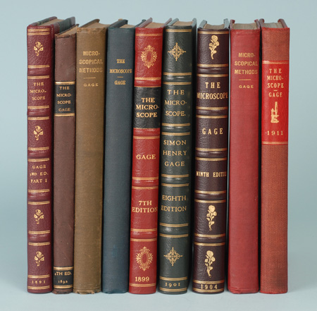
The 1891 third edition of Gage’s book was now hardbound, titled The Microscope and Histology, and described as being “entirely re-written”. As in earlier versions, microscopical methods were described in Part 1, and histological methods were described in Part 2. Part 2 is rarely seen, probably because blank pages were provided for student drawings, and these personalized productions are less likely to have survived. Figure 6a, far left, illustrates the third edition’s Part 1, The Microscope and Microscopical Methods (GAGE 1891).There are 96 individually numbered pages; the right-hand page being blank. This particular copy belonged to W. A. Turnbull, M. D., with his ownership label dated February 12, 1894. He left in a publisher’s note dated October 23, 1891, which described the book as containing five full page plates and eight text figures; the price bound in heavy manila was $1.00; half leather $1.25. For this edition, Gage introduces new chapters on the Micro-Spectroscope, the Micro-Polariscope, and interestingly, introduces experiments in Micro-Chemistry, using chemicals suggested by Professor Dennis of the Chemical Department; Herapath’s method of determining minute quantities of quinine is included.
Gage’s fourth edition (Figure 6) was published by James W. Queen & Co., Philadelphia in 1892 (GAGE 1892); the covers are semi-flexible. Gage adds an additional plate on the micro-spectroscope and micro-polariscope to this edition, and makes minor changes.
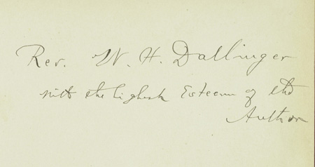
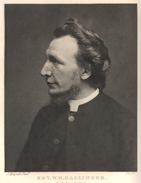
The fifth edition of Gage’s book (GAGE 1894) was rewritten, enlarged to 165 pages, and illustrated with 103 text figures; the enlargement is due not only to elaboration of the matter in the previous edition, but by the addition of a wholly new chapter on photomicrography used in photographing natural history objects—Gage’s editions are characterized by the addition of new chapters on specialized topics. The pages are again printed almost exclusively on the left-hand page; the right-hand page throughout is blank. The fifth edition is found in both brown and in red cloth. The particular copy of the fifth edition in hand here is special, in that it is known as an “association copy”; Gage’s handwritten presentation (Figure 7) reads: “Rev. W. H. Dallinger/with the highest Esteem of the/Author. The Reverend W. H. Dallinger (Figure 8) was, of course, the editor of the last two editions of Carpenter’s The Microscope and Its Revelations, the President of the Royal Microscopical Society, etc.
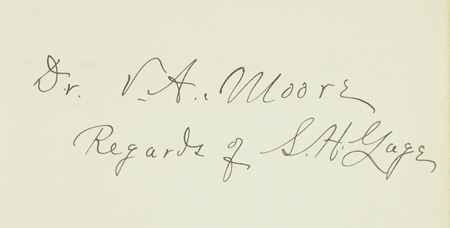
The sixth edition of Gage’s book (GAGE 1896b) is expanded to 237 pages and 165 text figures, with the addition of an Appendix on the use of Abbe’s Test Plate and Apertometer, and the preparation of diagrams and drawings for photo-engraving. Each page is now printed; the blank pages are no longer supplied. Illustrations of the latest microscopes, both domestic and imported, are abundant—another characteristic of Gage is the updating of instruments with each new edition. Gage’s title as Professor of Microscopy, Histology and Embryology now also indicates his affiliation with the New York State Veterinary College, in addition to Cornell University. This particular copy of the sixth edition was a presentation copy to Dr. V. A. Moore of Cornell’s Veterinary School, and has Gage’s signed holograph inscription (Figure 9).
The seventh edition of Gage’s book (GAGE 1899) (Figure 6), three years after the sixth, is essentially unchanged, with the exception of minor rewriting of a half dozen sections, and the addition of more advantageous methods in some sections.
The eighth edition The Microscope (GAGE 1901) (Figure 6) is expanded now to 299 pages, and is illustrated by over two hundred figures. In this edition, the chapters have all been recast, new figures added, the matter increased, and there is a new chapter on classroom demonstrations with the microscope using the projection microscope.
In the ninth edition (GAGE 1904) important changes are made in those parts having to do with serial sections, and to microchemistry; the chapter on the projection microscope is entirely rewritten; and all of the illustrations of microscopes and apparatus are updated.
Gage retired in 1908 at the age of 57, and the tenth edition (GAGE 1908) of his book came out the same year, with his title now indicated as Professor Emeritus. The 359-page book now has 258 illustrations. His son, Henry Phelps Gage, is given credit for help in optics, microchemistry, and photography, and his wife, Susanna Phelps Gage, is acknowledged for critical revision of the whole work, for proof reading, and for preparation of the index. Gage saves final thanks for Professor Burt Green Wilder “who encouraged me when a student to undertake work with the microscope, and gave me every facility in his power.” Gage dedicates this edition to his students and “to the Unknown Groups of Workers who have received aid from earlier editions…….”
The eleventh edition (GAGE 1911) (Figure 6) is practically unchanged from the 1908 tenth edition; a few typographical errors are indicated, and six pages of additions are included.
There are a number of important changes in the 1917 twelfth edition (GAGE 1917) (Figure 10) of Gage’s book. Having been retired, Gage now has the time to increase the contents to 472 pages and 252 figures, while at the same time omitting entirely the sections on microchemistry and metallography. It is interesting to pause here a moment, and consider the reason for Gage to have eliminated microchemistry and metallography.
In fact, he tells us in the twelfth edition Preface that it was eliminated “because the Micro-Chemistry of Dr. E. M. Chamot, which has recently appeared treats these and indeed all matters pertaining to the chemico-physical side of microscopy in an adequate manner.” What Gage is referring to is Emile Monnin Chamot’s Elementary Chemical Microscopy (CHAMOT 1916) published the previous year. Chamot (Figure 11) was by then Professor of Sanitary Chemistry and Toxicology at Cornell, and, what is little known amongst practicing microchemists, is that Chamot was influenced by Gage, or, to put it in Chamot’s own words from the Preface of his book on chemical microscopy “To Simon Henry Gage, Professor Emeritus of Histology and Embryology, the writer also acknowledges his indebtedness for much that is here presented. It is largely due to the spirit of optimism and love for research with which this indefatigable investigator is ever surrounded that the author was originally led to enter the field of applied microscopy when first a student.”
This twelfth edition also has a new chapter devoted to the history of lenses and the microscope. Another thing that needs to be mentioned about the 1917 twelfth edition is that Gage’s wife, Susanna, had died two years earlier, and this edition is touchingly dedicated to her: “To Susanna Phelps Gage, who as a student received help and inspiration from the earliest edition of this work, and who aided in every way possible by drawing, index-making and literary criticism in improving the later editions; and who now has entered into the possibilities of eternity….”
In 1918, at the age of 67, Gage came out of retirement to relieve the shortage of teachers due to the First World War. Two years later, the thirteenth edition (GAGE 1920a) of his book appeared; it was now 474 pages, with 275 figures (Figure 10). In this edition there is added emphasis on darkfield microscopy, some new apparatus, and Corning’s Daylight Glass. Gage also calls attention to a Symposium on the Microscope; its design, construction, and application, held by the Royal Microscopical Society, the Faraday Society, the Optical Society, and the Photographic Society that was held in the rooms of the Royal Society in January, 1920.
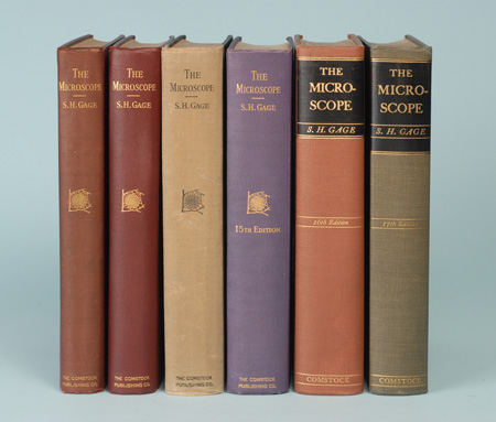
There were four illustrious editions of Gage’s book yet to come, two of which were perhaps best known by name as the Dark-Field edition, incorporating a black binding, and the Ultraviolet edition, in a violet binding (Figure 12). The 1925 fourteenth edition of Gage’s book (GAGE 1925) had a second printing in 1927. Gage was 74 when this 517-page edition came out. As the name suggests, this new edition stresses the darkfield technique, but new, stronger electric light sources in addition to daylight filtration are also discussed. The greater use of binocular heads is noted and discussed. The chapter on the history of microscopes has a plate of portraits of prominent figures in the history of lenses and microscopes. This edition is dedicated to Gage’s sister, Mary, a physician. This fourteenth Dark-Field edition of 1925 should perhaps have been anticipated, since five years earlier Gage had already published an extensive article called, Modern Dark-Field Microscopy and the History of its Development, in the Transactions of the American Microscopical Society (GAGE 1920b).
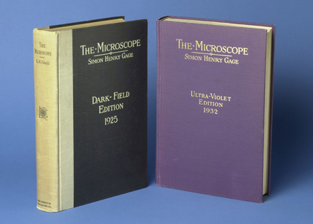
At age 81, Gage had published the fifteenth edition of his work, now simply titled, The Microscope (GAGE 1932), easily recognized by its name, the Ultra-Violet Edition 1932, in its violet cloth binding. It is now 589 pages, with 1 plate and 291 figures. In addition to the general revisions and rewriting, there is, of course, a wholly new chapter on ultraviolet radiation in microscopy. Importantly, special emphasis is now being placed on what was called “the physical analysis of structure;” that is, the integration of the polarizing microscope, the microspectroscope, darkfield, and ultraviolet. In mentioning the new differential staining methods, Gage recalls the limitations on the older histologists of the last century who, with only two or three stains still found out the most fundamental things in the structure of plants and animals, and laid the foundation for all of the advances made since. Gage knew of this firsthand, as he was now 81, and had lived through it all. He acknowledges help now from Chamot, and from his son Dr. Henry Phelps Gage. Gage dedicates this Ultra-Violet edition to both Cornell University, and to Cornell’s President, Livingston Farrand.
Chamot was right—Gage is indefatigable! At age 85 he produces the sixteenth edition of The Microscope (GAGE 1936) (Figure 10). It is now 615 pages, with 313 figures. In addition to rewriting and updating all instrument figures, Gage added a new chapter on Micro-Incineration. Reflected ultraviolet light is now also possible through the use of aluminized mirrors. This edition is dedicated to Theobald Smith “…pupil, friend, and master investigator who opened new paths to the human mind.” Gage acknowledges the help of Clara Covert Starrett for this edition; they were married three years earlier.
Incredibly, Gage, now 90 years old, produces the seventeenth, and last, edition of The Microscope (GAGE 1941) (Figure 10). Gage makes changes in every chapter, in both text and illustrations. For this edition, he calls attention to the newly devised electron microscope, the use of Polaroid for the micropolariscope, new plastic mounting media in place of Canada balsam, and high pressure mercury vapor lamps for ultraviolet radiation and bright mercury lines for photomicrography. He acknowledges the help of his wife, Clara Starrett Gage, Ph.D. in preparing illustrations, making corrections, and revising the index.
There was a second printing of this edition in 1943. Gage died the following year at age 93.
Optic Projection
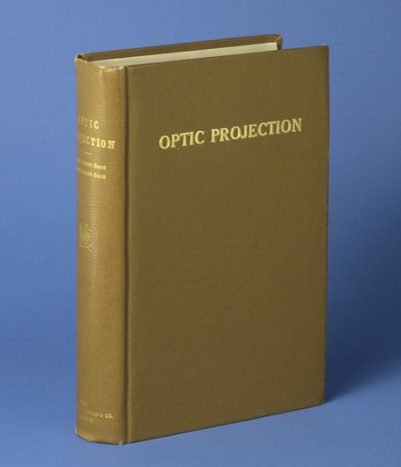
Between the 1911 eleventh edition and 1917 twelfth edition of his book on the microscope, Gage coauthored a book with his son. This book, Optic Projection (GAGE and GAGE 1914) (Figure 13) was devoted to the principles, installation, and use of the magic lantern, the forerunner of the modern slide projector and digital imaging; the projection microscope (still in use in schools as the microprojector); the reflecting lantern (today’s opaque projector); and the moving picture machine (today’s film projector). Gage wrote ten of the chapters in this 731-page book, and his son wrote five of the chapters. Dolbear (DOLBEAR 1887) and Wright (WRIGHT 1891) had both written books on this topic, but that of Gage and Gage brought the subject up to date. Gage used these projection methods extensively in his classes, and it was natural for him to want to share those things which he had learned.
Microscopy in America
Gage had started a history of lenses and the microscope in the form of a chapter which he introduced in the twelfth edition of The Microscope, but he intended to write a more extensive history, especially as it applied to microscopy in America from 1830 to 1945. He was engaged in the writing of this history and it was almost finished when he died. Gage’s wife Clara and his son Dr. Henry Phelps Gage asked Oscar Richards to complete the history. Richards did the editing with minimal change of text, largely attempting a final organization suggested by Gage’s notes. Gage’s Microscopy in America (1830-1945) was finally published by Richards as a Supplement to the Transactions of the American Microscopical Society (RICHARDS 1964).
Conclusion
Simon Henry Gage was, clearly, a remarkable, prolific, and influential figure in American microscopy. His writings are still fruitful to the modern practicing microscopist, if they only take the time to read them. He lived the history of the subject he wrote about. Incidentally, his actual brain is on display, preserved in formalin and stored in a glass jar, along with that of Dr. Burt Green Wilder, Cornell’s first zoologist, on the second floor of Uris Hall at Cornell University—part of a brain collection that once numbered 1,600 animal and human brains—but that is another story.
Acknowledgements
I am deeply grateful to Amanda Mott, Administrative Assistant, External Relations & Marketing, Cornell University, College of Veterinary Medicine, for arranging Cornell’s College of Veterinary Medicine Archives/Image Lab to supply a digital photo of the magnificent portrait of Professor Simon Henry Gage that currently hangs in Cornell’s Flower-Sprecher Veterinary Library.
References
CHAMOT, E. M. (1916) Elementary Chemical Microscopy. John Wiley & Sons, New York and Chapman & Hall, London, 410 p.
DOLBEAR, A.E. (1887) The Art of Projecting; A Manual of Experimentation in Physics, Chemistry, and Natural History with the Porte Lumiere and Magic Lantern. New Edition (First, 1877). Lothrop, Lee & Shepard Co., Boston, 178 + p.
GAGE, S. H. (1880-1881) Microscopical Technology. The “First Edition” of Gage’s book on the microscope printed as student handouts by the hectograph method. Ithaca, New York, 54 p.
GAGE, S. H. (1885-1886) Notes on Histological Methods, Second edition. Andrus & Church, Ithaca, New York, 56 p.
GAGE, S. H. (1887) Notes on Histological Methods, For the Use of Students of the Anatomical Department of Cornell University, Second edition. Andrus & Church, Ithaca, New York, 32 p. [Kessinger reprint available].
GAGE, S. H. (1891) The Microscope and Histology; Part I The Microscope and Microscopical Methods, Third edition. Andrus & Church, Ithaca, New York, 96 p.
GAGE, S. H. (1892) The Microscope and Histology; Part I The Microscope and Microscopical Methods, Fourth edition. James W. Queen & Co., Philadelphia, Pennsylvania, 96 p.
GAGE, S. H. (1893) The Lake and Brook Lampreys of New York; Especially Those of Cayuga and Seneca Lakes. Reprinted from The Wilder Quarter-Century Book, 421-493 + maps and plates.
GAGE, S. H. (1894) The Microscope and Histology; Part I The Microscope and Microscopical Methods, Fifth edition. Comstock Publishing Co., Ithaca, New York, 165 p.
GAGE, S. H. (1896a) The Processes of Life Revealed by the Microscope; A Plea for Physiological Histology. Transactions of the American Microscopical Society 17 3-29. Also, Smithsonian Institution Report 381-396 (1896). Printed almost entirely in Science n.s. 2 209-218 (1895) and in American Monthly Microscopical Journal 16, 292-311 (1895).
GAGE, S. H. (1896b) The Microscope and Microscopical Methods, Sixth edition. Comstock Publishing Co., Ithaca, New York, 237 p.
GAGE, S. H. (1899) The Microscope; An Introduction to Microscopic Methods and to Histology, Seventh edition revised. Comstock Publishing Co., Ithaca, New York, 237 p.
GAGE, S. H. (1901) The Microscope; An Introduction to Microscopic Methods and to Histology, Eighth edition. Comstock Publishing Co., Ithaca, New York, 299 p.
GAGE, S. H. (1904) The Microscope; An Introduction to Microscopic Methods and to Histology, Ninth edition. Comstock Publishing Co., Ithaca, New York, 299 p.
GAGE, S. H. (1908) The Microscope; An Introduction to Microscopic Methods and to Histology, Tenth edition. Comstock Publishing Co., Ithaca, New York, 359 p.
GAGE, S. H. (1911) The Microscope; An Introduction to Microscopic Methods and to Histology, 11th edition. Comstock Publishing Co., Ithaca, New York, 359 p.
GAGE, S. H. (1917) The Microscope; An Introduction to Microscopic Methods and to Histology, Twelfth edition. Comstock Publishing Co., Ithaca, New York, 472 p.
GAGE, S. H. (1920a) The Microscope; An Introduction to Microscopic Methods and to Histology, Thirteenth edition. Comstock Publishing Co., Ithaca, New York, 474 p.
GAGE, S. H. (1920b) Modern Dark-Field Microscopy and the History of its Development. Transactions of the American Microscopical Society XXXIX (2) 95-141.
GAGE, S. H. (1925) The Microscope; An Introduction to Microscopic Methods and to Histology, Darkfield (14th) edition. Comstock Publishing Co., Ithaca, New York, 517 p. Second Printing 1927.
GAGE, S. H. (1932) The Microscope, Ultra-Violet (15th) edition. Comstock Publishing Co., Ithaca, New York, 589 p.
GAGE, S. H. (1936) The Microscope, Sixteenth edition. Comstock Publishing Co., Ithaca, New York, 615 p.
GAGE, S. H. (1941) The Microscope, Seventeenth edition. Comstock Publishing Co., Ithaca, New York, 617 p. Second Printing 1943.
GAGE, S. H. and GAGE, H. P. (1914) Optic Projection; Principles, Installation and Use of the Magic Lantern, Projection Microscope, Reflecting Lantern, Moving Picture Machine. Comstock Publishing Co., Ithaca, New York, 731 p.
Journal of Applied Microscopy (1898). Gage’s portrait appears on page 161, Volume 1, Number 9, September, 1898. The caption reads: “Prof. S. H. Gage, Cornell University, Member Executive Committee, A. M. S., 1898-9.”
RICHARDS, O. W., editor (1964). Microscopy in America (1830-1945) By Simon Henry Gage. Transactions of the American Microscopical Society, LXXXIII, No. 4, Supplement, October 1964, 125 p.
WRIGHT, L. (1891) Optical Projection; A Treatise on the Use of the Lantern in Exhibition and Scientific Demonstration, Second edition. Longmans, Green, and Co., London and New York, 427 p.
![]()
Comments
add comment